Case Study
A 37-year-old male visited a dentist for a routine checkup. Oral examination revealed a radiopacity in the maxillary sinus.
History
When questioned about the sinus area, the patient denied any history of symptoms associated with this region. The patient appeared to be in a general good state of health, with no significant medical history. The patient's dental history included regular examinations and routine treatment. At the time of the dental appointment, the patient was not taking any medications.
Examinations
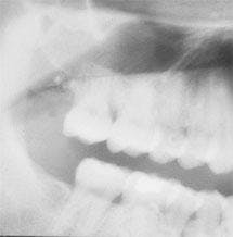
The patient's vital signs were found to be within normal limits. Examination of the head and neck region revealed no enlarged or palpable lymph nodes. Examination of the soft tissues of the oral cavity revealed no unusual findings. Radiographic examination revealed a large radiopacity projecting from the floor of the left maxillary sinus (see radiograph). The radiopacity appeared as a smooth, dome-shaped, homogeneous mass. The teeth adjacent to the left maxillary sinus were tested for vitality; all tested vital.
Clinical diagnosis
Based on the clinical and radiographic information available, which one of the following is the most likely diagnosis?
- periapical cyst
- malignant tumor
- sinusitis
- antral pseudocyst
- periapical abscess
Diagnosis
- antral pseudocyst
Discussion
The antral pseudocyst is a common and well-documented finding on a panoramic radiograph. Most are discovered during routine radiographic examination and have little clinical significance. The antral pseudocyst should not be confused with the mucocele of the sinus, a destructive lesion of the sinus that requires surgical intervention.
The antral pseudocyst is believed to be caused by an inflammatory exudate that accumulates under the maxillary sinus mucosa and results in a sessile elevation. The exudate is serous (fluid-like) and not mucous in nature. This fluid is found below the periosteum of the floor of the maxillary sinus and results in the separation and elevation of the sinus mucosal lining from the bony floor. The cause of the fluid accumulation that results in the antral pseudocyst is unclear, although some studies suggest an adjacent odontogenic infection may precipitate the formation of this lesion. A primary irritation of the sinus lining is also considered a possible cause for the antral pseudocyst.
Clinical features
The antral pseudocyst is estimated to occur in 1.5 to 10 percent of the population. The antral pseudocyst of the maxillary sinus can be seen in any age group. Males are affected more frequently than females. The antral pseudocyst is asymptomatic. In contrast, the true sinus mucocele — a lesion that is capable of expanding and destroying bone — is associated with symptoms such as pain and tenderness in the areas of the teeth and face adjacent to the sinus.
The antral pseudocyst may vary in size from a small dome-shaped mass to a very large dome-shaped lesion that fills the entire sinus cavity. Over time, the antral pseudocyst may remain stationary, increase or decrease in size, or disappear for no apparent reason. The antral pseudocyst is best demonstrated on a panoramic film and is often discovered during routine radiographic examination. It may also be seen on maxillary posterior periapical films or extraoral films such as the Water's and the posterior-anterior skull films.
Radiographically, the antral pseudocyst appears as a dome-shaped radiopacity extending from the floor of the maxillary sinus. The lesion appears as a homogeneous, soft-tissue radiopacity. The borders of the lesion exhibit the same density as the body of the mass and normal anatomic landmarks can be seen through the image of the mucous retention cyst. The antral pseudocyst most often occurs unilaterally, but may also be seen bilaterally.
Diagnosis
The location and radiographic appearance of the antral pseudocyst are considered to be diagnostic. Based on the radiographic appearance, there are a number of lesions that may be confused with the antral pseudocyst, including tooth-related cysts, the sinus mucocele, and malignant tumors of the sinus.
Treatment
The antral pseudocyst usually persists unchanged or disappears for no apparent reason. No treatment is necessary because the lesion is limited in growth and not destructive. The teeth adjacent to the antral pseudocyst should be thoroughly evaluated and any areas of odontogenic infection should be eliminated.
If the diagnosis is questionable, or if symptoms are present, the patient should be referred to an ear, nose & throat specialist for evaluation. The patient with an antral pseudocyst should be informed that the lesion is present and be reassured concerning the benign nature of the lesion. Because the majority of antral pseudocysts regress spontaneously, periodic radiographic examination with panoramic films can be used to follow the lesion.
Joen Iannucci Haring, DDS, MS, is an associate professor of clinical dentistry, Section of Primary Care, The Ohio State University College of Dentistry.

