When anesthetizing a quadrant is necessary, a maxillary nerve block may be appropriate.

Dental hygienists should periodically review the maxillary nerve, its branches, and techniques available to achieve anesthesia of the maxilla. It’s no secret that, prior to root planing, many people dread the injections more than the procedure. By offering an alternative that reduces the number of injections, both clients and practitioners can look forward to a less stressful procedure.
Anesthesia of the maxillary division of the trigeminal nerve is a well-known procedure described by Malamed that can use either of two methods. In one technique, the practitioner must find the greater palatine foramen and pass the needle through the greater palatine canal. This is difficult, and occasionally the canal is impassable. The other technique described by Malamed is easier and calls for advancing the needle posterior and superior to the maxillary tuberosity. While the latter technique may be easier, there is a higher risk of puncturing the pterygoid venous plexus or maxillary artery in the pterygomaxillary fossa. In both cases, for an average size adult the needle must pass through 30 mm of soft tissue to reach its destination in the pterygomaxillary fossa.
Dental anesthesia for maxillary structures is typically learned by anesthetizing each of the branches distal to their separation from the maxillary nerve. If the maxillary nerve is not anesthetized before it branches off, as many as five nerve blocks may be needed to achieve thorough anesthesia of a maxillary quadrant. These blocks are:
• Posterior superior alveolar nerve (injected near the posterior surface of the maxilla)
• Middle superior alveolar nerve (injected above the second premolar)
• Anterior superior alveolar nerve (injected at the infraorbital foramen above the canine or first premolar)
• Greater palatine nerve (injected at the greater palatine foramen medial to the second molar)
• Nasopalatine nerve (injected at the incisive foramen, behind the central incisors)
In many cases, anesthetizing an entire quadrant is unnecessary and may seem excessive to a client who is having only one or two teeth treated. It is possible to anesthetize a smaller area with sufficient depth and duration. In these cases, nerve block anesthesia in one of the branches of the maxillary nerve is appropriate.
Also, many practitioners are afraid to use a maxillary nerve block due to the proximity of the nerve to blood vessels in the pterygopalatine fossa. Yet, after several experiences of achieving significant anesthesia of several maxillary nerve branches following administration of anesthetics intended for the posterior superior alveolar nerve block, I wonder about the safety of achieving sufficient analgesia of oral structures innervated by the maxillary nerve block.
If we can do this, we can save time and trauma to clients by using fewer injections and less anesthetic. This is particularly important to clients at high risk of adverse reaction to local anesthetics. If the maxillary block fails to achieve sufficient analgesia, areas that need additional injections will receive them at the first attempt to anesthetize the tissues through blocks of the maxillary nerve branches. The injection intended for the maxillary nerve will provide anesthesia for the posterior superior alveolar nerve.
Sensory neuron cell bodies of the trigeminal nerve are in the trigeminal ganglion. This ganglion is known by two other names - the Gasserian ganglion and the semilunar ganglion (due to its crescent shape). The trigeminal ganglion is in a pouch of dura mater known as the trigeminal cave or Meckel’s cave, which is located in a depression on the petrous portion of the temporal bone within the middle cranial fossa.
Sensory and motor nuclei and cell bodies of proprioceptive nerves of the trigeminal nerve are in the pons. The sensory and motor nuclei of the trigeminal nerve are at a level below the superior cerebellar peduncle to the level of the middle cerebellar peduncle, and the nerve exits the pons near the middle cerebellar peduncle. Nerve cells that form synapses in the sensory nuclei transmit tactile, temperature, and pain sensations.
The trigeminal nerve is recognized as three distinct divisions - ophthalmic (V1), maxillary (V2), and mandibular (V3). Each division of the trigeminal nerve passes through a different foramen to leave the skull and innervate its designated location. V1 passes through the superior orbital fissure, and V3 through the foramen ovale. After it provides a middle meningeal nerve to innervate the dura mater and passes anteriorly from the trigeminal ganglion, V2 passes through the foramen rotundum within the greater wing of the sphenoid bone. At that point, V2 is in the most superior part of the pterygopalatine fossa. It passes anteriorly between the sphenoid and palatine bones, then laterally along the posterior surface of the maxilla. While in the pterygopalatine fossa, V2 has branches to the sphenopalatine ganglion and the zygomatic and posterior superior alveolar nerves. Nerves from the sphenopalatine ganglion innervate the orbit of the eye, the hard and soft palate, and parts of the nasal cavity, palatine tonsil, nasopharynx, and ethmoid sinuses. The zygomatic nerve goes further into the zygomaticofacial and zygomaticotemporal nerves, and supplies skin over the zygomatic process and anterior part of the temple.
The maxillary nerve continues through the inferior orbital fissure, at which point it becomes the infraorbital nerve, continues along the infraorbital groove, and reaches the face through the infraorbital canal, which ends at the infraorbital foramen. The trigeminal nerve carries branchial motor nerves originating from the pons; however, those efferent nerves travel along V3. Visceral motor nerves originating from other cranial nerves travel with all divisions of the trigeminal nerve, but are not considered part of the trigeminal nerve.
The maxillary nerve has several branches that play a major role in pain management during oral health care procedures.
• Posterior superior alveolar nerve (branches off in the pterygopalatine fossa, runs inferiorly along the posterior border of the maxilla, and passes through that surface to innervate the maxillary sinus, molars, buccal gingiva, and cheek)
• Middle superior alveolar nerve (branches off from the infraorbital nerve within the infraorbital fissure, passes lateral to the maxillary sinus, and innervates the first molar, premolars, buccal gingiva, and cheek)
• Anterior superior alveolar nerve (branches off from the infraorbital nerve in the infraorbital canal, passes anterior to the maxillary sinus, and innervates the anterior teeth, labial gingiva, lip, and parts of the nose)
• Ganglionic branches (pass through the pterygopalatine ganglion to innervate the palate)
Bergman, Afifi, and Miyauchi describe some variations of the maxillary nerve. These include:
• Missing middle superior alveolar nerve
• Separate nerve branch parallel to the infraorbital nerve supplying the upper lip
• Bifid maxillary nerve
• Posterior superior alveolar nerve innervating areas normally covered by the long buccal nerve
• Branches from the sphenopalatine ganglion supplying the abducens, optic, or ciliary nerves
• Various exchanges of nerve coverage among zygomaticofacial, zygomaticotemporal, infraorbital, and lacrimal nerves
Dental hygienists want a comfortable setting in which to perform their procedures. At the same time, many clients are not accustomed to the idea that a dental hygiene procedure might need to be uncomfortable to be effective. Multiple injections for pain control during a hygiene visit may be a surprise to someone whose experiences with dental hygienists have not addressed periodontal disease. Offering the smallest number of injections necessary for proper treatment helps the dental hygienist maintain rapport, keep clients on their maintenance schedules, and keep the lines of communication open to educate clients about treatment and home care.
Howard M Notgarnie, RDH, MA, practices dental hygiene in Colorado, and has eight years experience in official positions in dental hygiene associations at the state and local levels.
References
Anderson, J. E. (1983). Grant’s Atlas of Anatomy, 8th ed. Baltimore: Williams & Wilkins.
Bergman, R. A., Afifi, A. K., & Miyauchi, R. (2005). Illustrated Encyclopedia of Human Anatomic Variation. Iowa City, IA: University of Iowa. Retrieved June 25, 2005 on http://www.vh.org/adult/provider/anatomy/AnatomicVariants/NervousSystem/Alphabetical/T.html
Malamed, S. F. (1997). Handbook of Local Anesthesia, 4th ed. St Louis: Mosby.
Snell, R. S. (1981). Clinical Anatomy for Medical Students, 2nd ed. Boston: Little, Brown & Company.
University of Kansas. (1996). Neuroanatomy Exam Study Sheet. Lawrence, KS: University of Kansas Retrieved June 26, 2005 on http://www.geocities.com/doctor_uae/neuro2.htm
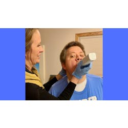
The following technique has been successful at achieving sufficient analgesia with few instances of positive aspiration and no instances of hematoma.
▲ Apply topical anesthetic to the area of intended initial penetration of the needle
▲ Assemble syringe with the chosen anesthetic and a long (32 mm) needle
▲ Identify landmarks: alveolar mucosa at the maxillary mucobuccal fold, buccal surface of maxilla, zygomatic process of maxilla
▲ Open the client’s mouth no more than half way and have the mandible in lateral excursion to the side of the injection. Additional improvement to access can be achieved by having the client turn his/her head to the side opposite the injection site
▲ Palpate the posterior surface of the zygomatic process where it meets the buccal surface of the maxilla using the index finger of the non-injecting hand
▲ Slide the finger posteriorly and laterally from the zygomatic process until a depression in the alveolar mucosa is visible between the finger and zygomatic process
▲ In a straight path, direct the needle in a posterior, superior, and medial direction, maintaining its position as close as possible to the posterior surface of the maxilla
▲ Penetrate to about one-half the length of the long needle (16 mm)
▲ Confirm negative aspiration
▲ Slowly inject 1.8 ml of anesthetic solution
▲ Remove the needle along the same path
This technique is consistent with Malamed’s description of the posterior superior alveolar nerve block. The use of a long needle allows better visualization of the angle at which the needle advances.
I personally tabulated 46 attempts at V2 anesthesia using this technique during a four-month period. Of these attempts, 15 (33 percent) resulted in profound anesthesia throughout the quadrant and 30 (65 percent) had sufficient analgesia and needed no further injections or topical anesthetic during root planing.
Of the clients who needed more anesthesia, all required injections at the anterior superior alveolar nerve, two required additional nasopalatine anesthesia, and one required additional greater palatine anesthesia. There were four (8.7 percent) positive aspirations with no hematomas. Two of those with positive aspiration and repositioning of the needle required injections at additional sites.
Interestingly, when performing root planing on maxillary and mandibular quadrants on the same visit and the same side, with one needle penetration site for inferior alveolar and lingual blocks and one needle penetration intended for V2, there were no instances in which the buccal gingiva of the mandibular molars required a separate injection. As the long buccal nerve via the anterior division of the mandibular nerve innervates this area, sufficient anesthetic diffuses to that nerve where it passes by the lateral pterygoid muscle.
Why does this technique provide sufficient analgesia for root planing in a majority of cases despite the deposition of anesthetic closer to the posterior superior alveolar nerve rather than the maxillary nerve?
I believe there are two explanations. First, the pterygopalatine fossa is a narrow space almost completely enclosed by the sphenoid, palatine, and maxillary bones. The dense periosteum surrounding the fossa is probably more resistant to diffusion of the anesthetic solution than the soft tissue within the fossa. It acts as a funnel, guiding movement of the fluid medially and superiorly toward the maxillary nerve until pressure from the solution has equilibrated with the surrounding tissue.
Second, although the funneling effect does not usually reach the levels of anesthetic needed to achieve profound anesthesia, the anesthetic significantly affects the narrower and unmyelinated nerves involved in pain sensation.
Given the presence of the pterygoid venous plexus in the pterygopalatine fossa, particular care must be used when selecting clients and administering anesthetics for the maxillary nerve block. An inexperienced operator should avoid a V2 block until he or she has successfully administered many posterior superior alveolar nerve blocks.
Additional contraindications to the technique described are a client with a small skull, such as a child, an uncooperative client, inflammation, and the risk of excessive bleeding.