by Ann M. Drewenski, RDH
Dental implants are a major investment, and with proper care can last a lifetime. Advantages of implants include comfort, heightened self-esteem, ease of eating, and improved appearance, speech, and oral health. With comprehensive education by the entire dental team, patients can better understand the importance of home care and regular maintenance visits.
Infection control lies at the heart of peri-implant treatment. With regular evaluation, plaque and calculus removal, and patient commitment to an infection-free environment, an implant can last a lifetime. As dental hygienists, we are obligated to educate and motivate every patient. In doing so, it’s imperative that we take into consideration each patient’s home care, systemic health, and periodontal history.
In order to maximize the longevity of dental implants, the clinician must understand the morphology of the peri-implant mucosa, the attachment between the mucosa and the titanium implant. It comprises the junctional epithelium, about 2 mm high, and the connective tissue zone of greater than or equal to 1 mm in height. This is the zone that protects the osseointegrated surface from environmental factors, such as plaque in the oral cavity. Proper monitoring and maintenance of this zone is essential to the health and longevity of a dental implant.
In 1991, Dr. Tord Berglundh, professor in the department of periodontology at the University of Gothenburg and the head of the periodontal research laboratory, studied the quality and composition of tissues around teeth and implants. The tooth surface consisted of cementum with bundles of fibers projecting radially in all directions. The implant surface demonstrated no cementum, and the fiber bundles ran parallel to the bone crest or were aligned in coarse bundles parallel to the bone surface. The composition of the connective tissue and the distribution of vascular structures in the compartment apical to the junctional epithelium were also different.
In 1992, Berglundh and Ingvar Ericsson, a dentist in Göteborg and a consultant for Nobel Biocare, studied the reaction of gingival and peri-implant mucosa to three-week and three-month plaque accumulation. They found that plaque formation and soft tissue inflammation was similar in gingival and peri-implant mucosa. However, due to a lack of fibroblasts, there was an increase in the duration of inflammation, and it progressed apically around the implant. Hence, bacterial plaque not only leads to gingivitis and periodontitis, but also the development of peri-implantitis.
Proper monitoring and maintenance is essential to ensure the durability and health of a dental implant. The key to long-term maintenance is to begin with a customized treatment plan for each patient. Oral hygiene instructions preoperatively will greatly benefit the patient and enhance the long-term predictability of the implant. The goal is to maintain a stable, infection-free, healthy environment.
Before the surgical procedure, the clinician should make sure there is no sign of infection and should have a current full mouth set of radiographs, full mouth periodontal (6 point) probe readings charted, a comprehensive exam, and a prophylaxis. Periodontal therapy, including scaling and root planing (four quadrants if required) or localized periodontal therapy (one to three teeth), laser therapy, Jet Polisher 2000, and adjunctive therapy (i.e., antibiotics placed at sites as needed), should be performed if needed. Chlorexadine gluconate (CHX 0.12%) and oral hygiene instruction should also be provided.
Personal hygiene must take place before, during, and after placement of dental implants. Prescription rinses of CHX should be used routinely. Chlorhexidine is an ADA and FDA approved, substantive, non-selective topical antibiotic with no current resistant strains of bacteria. CHX can reduce plaque and gingivitis up to 55%. There are a multitude of other dental hygiene products on the market, and since each patient has many different needs, the dental hygienist has a significant role in guiding the patient to whatever product works best for him or her. Whether the dental aid is an oral irrigator, interproximal toothbrush, flossing aid, electric toothbrush, or manual toothbrush, patients should be well educated on the products that will help them maintain a healthy and stable environment. This will not only benefit the dental implant, but can help patients maintain their systemic health since bacterial plaque has been found to be associated with coronary issues, respiratory problems, diabetes, pregnancy, and low birth-weight babies.
During the maintenance visit, and after the implants have been placed, the dental hygienist should put all his or her skills and education into action by assessing and evaluating the peri-implant tissue margin, implant body, prosthetic abutment-to-implant collar connection, and prosthesis.
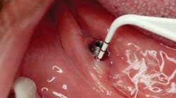
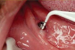
Clinical inspection for signs of inflammation, exudates, mobility, increased sulcus depth, and occlusal trauma are very important to evaluating the status and durability of the dental implant or prosthesis. Clinical assessments used routinely, with the exception of periodontal probing around the peri-implant, should be documented. Routine periodontal probe readings are not recommended. Probe readings should only be used when needed due to signs of inflammation, bleeding, or loose implant. Routine probing could cause damage to the weak epithelial attachment around the implant, possibly creating a pathway for periodontal pathogens. Use of a plastic rather than metal probe is recommended should probing be indicated (see Figure 1).
A three-month maintenance schedule should be implemented during the first year after the implant is restored, especially if the patient has lost teeth due to periodontal disease or medical conditions associated with periodontal disease. After 12 months, if the implants are stable, the peri-implant tissues are healthy (no signs of infection, inflammation, bleeding on scaling, or occlusal trauma), and an infection-free environment has been maintained, a four-month regimen can be implemented.
Radiographs are taken every 12 to 18 months (during maintenance visits) for any signs of cement, calculus, or interference with stability and osseointegration of the implant. Long-term evaluation is needed because the peri-implant mucosal seal may be a less effective barrier to bacterial plaque than the periodontal attachment around the tooth. There is also less vascularity in the gingival tissue surrounding the dental implant compared to the tooth.
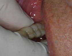
The dental hygienist should assess and evaluate any significant changes in the color, contour, and consistency of the soft tissue. Mobility of a healthy implant should be non-existent when adequate supporting alveolar bone is present. (Illustration No. 3)
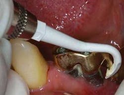
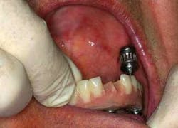
Great care and caution should be practiced when cleaning the dental implant. Any metal instruments can scratch the implant surface, leading to micro-roughness and gouging of the implant’s polished collar. Titanium dental implants can be polished using a rubber cup with a nonabrasive polish paste. Hand scalers for cleaning dental implants should be made of plastic, Teflon, or wood. An ultrasonic cleaning instrument can be used if there’s a shield over the tip. The Jetpolisher 2000 has a unique homogenous stream technology that does not create any abrasion on a titanium implant. (Illustration Nos. 4 and 5)
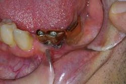
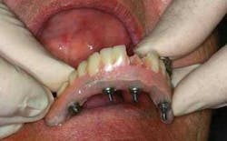
It’s necessary to remove a fixed prosthesis at least once every two years. Bacteria accumulate under the fixed prosthesis, causing infection and the possibility of losing the implants. This task can be completed and performed by a dental hygienist using a torque wrench and great caution. (Illustration No. 6)
In conclusion, the critical factors for longevity of an implant are maintenance by a professional, and patient understanding of the importance of keeping an infection-free environment. Implants are an investment and should result in a healthy, happy patient.
About the Author
Ann M. Drewenski, RDH, is a member of the American Academy of Cosmetic Dentistry and has conducted presentations on implant mainentenace and implant longevity in the Chicago area. She is on the board of trustees for the Chicago Component of the American Dental Hygienists’ Association.
