by Nancy W. Burkhart, RDH, EdD
[email protected]
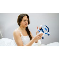
Your patient is a 45-year-old male who has complaints of tenderness in the region of No. 31 lingual with a shooting pain on the right side from mandible to the chin (see Figure 1). The patient was referred to the oral pathology department of your health facility months earlier with similar complaints. No pathology was found, and no clinical or radiographic findings were noted at the time. The patient was referred back to his general dentist. Several months later, the pain returned with an obvious lesion on the lingual area of No. 31. The patient reports a dull pain that is more pronounced when chewing. He is not aware of any trauma to the area in question.

Figure 1: Clinical slide of LMS depicting ulcerated lesion with evident bone fragment. Slide courtesy of Dr. Melissa Dennison, Bedford, N.H.
The diagnosis is lingual mandibular sequestration (LMS), which arises, as the name implies, in the mandibular molar region. The natural alignment of the teeth usually provides a sheltered environment, protecting the tissue from chronic assault and contamination from debris. When a tooth is extracted, the area is more vulnerable to irritation because of this loss of protection. Additionally, pressure from eating may cause a chronic type of inflammation and pressure within the tissues and the bone due to constant forces in the molar region, ultimately producing necrotic bone.
Osteonecrosis is also associated with several predisposing factors: dental extractions, trauma, systemic factors such as diabetes, immunosuppression, Paget's disease, osteopetrosis, and radiation therapy. Osteonecrosis associated with bisphosphonate therapy is also mentioned because the drugs suppress osteoclasts and inhibit the remodeling of bone. Bedogni, et al. (2010) report osteonecrosis associated with the use of alendronate (Fosamax), the development of lingual mandibular sequestration, and the subsequent failure of dental implants in some cases.
Patients describe the pain as one of a "dull ache" that often intensifies with chewing and swallowing. Ulceration may occur along with the appearance of osseous fragments. The tongue may become ulcerated from rubbing the bone fragments.
Eleven cases of lingual mandibular sequestration were documented by Peters and colleagues in 1993. It is suggested that the molar region may be more susceptible to this type of sequestration and that mild trauma may be enough to trigger the changes in bone and cause the ulceration. The radiograph presented in this case is typical with LMS because it appears normal and does not show radiographic changes, since the lesions involve only a limited area of the lingual cortical plate (see Figure 2).
Kessler (2005) suggests that there may be some peculiarity of the anatomic site that predisposes the mandibular lingual region of the mouth to the development of this type of lesion. He expands this theory to explain that the preponderance of cases is documented in the area of the mylohyoid line. The line represents the attachment of the lingual border of the mandible where the mylohyoid muscle inserts. He further explains that this line of muscle runs along the length of the mandible but is superiorly positioned in the second and third molar region. The position makes the area more vulnerable to trauma, ulceration, and fracture.

Figure 2: The periapical radiograph from this case is typical because LMS does not show radiographic changes, since the lesions involve only a limited area of the lingual cortical plate. Slide courtesy of Dr. Melissa Dennison, Bedford, N.H.
Kessler points out that "if the mylohyoid line is elevated and closer to the mucosal surface beneath the ulcer, microorganisms and resultant inflammation may more easily extend to involve the bony protuberance. This would produce a localized area of osteomyelitis with subsequent bone necrosis and sequestration."

Figure 3: The bone in the photomicrograph appears nonvital, and it has bacterial colonies adherent on its surface. Both features are typically seen in a sequestrum. Slide courtesy of Dr. Lynn Soloman, Tufts University School of Dental Medicine.
Small areas of bone begin to detach and the process of sequestration begins to occur. As osteoclasts attempt to resorb the bone fragments, inflammatory cells produce the heavy granulation tissue that occurs as the spicules of bone try to work their way to the surface of the tissue. The bone in the photomicrograph appears nonvital and it has bacterial colonies adherent on its surface – both features typically seen in a sequestrum (see Figure 3). The histology is certainly not specific since the lingual mandibular lesion is not really any different from a sequestrum anywhere else. It is just the consistency of the location that makes it a more distinctive presentation.
The patients presenting with lingual mandibular sequestration in the Peters report had a median age of 47 with an age range of 32-57. The length of time from onset to treatment was reported to be three weeks duration. Males outnumber females seven to four. This ratio is different for those found in bisphosphonate use since most individuals using these products are females.
On many occasions, it is possible to gently remove spicules of bone, resulting in complete healing. However, treatment usually involves surgical removal of the bone fragment, which normally results in healing of the affected area. The choice of therapy depends upon the severity of the symptoms and may include surgery, antibiotics, and chlorhexidine rinses.
As always, keep listening to your patients and always ask good questions!
References:
Bedogni A, Bettini G, Totola A, Saia G, Nocini PF. Oral bisphosphonate-associated osteonecrosis of the jaw after implant surgery: a case report and literature review. J Oral Maxillofac Surg. 2010 Jul:68(7):1662-6.
Jackson I, Malden N. Lingual mucosal ulceration with mandibular sequestration. Dent Update 2007 Nov:34(9): 573-7.
Kessler HP. Oral and maxillofacial pathology case of the month. Lingual mandibular sequestration with ulceration. Tex Dent J. 2005 Feb;122(2):198-9. 206-7.
Peters E, Lovas GL, Wysocki GP. Lingual mandibular sequestration and ulceration. Oral Surg Oral Med Oral Pathol 1993, 75:739-743.
Nancy W. Burkhart, BSDH, EdD, is an adjunct associate professor in the department of periodontics, Baylor College of Dentistry and the Texas A & M Health Science Center, Dallas. Dr. Burkhart is founder and co-host of the International Oral Lichen Planus Support Group http://www.bcd.tamhsc.edu/outreach/lichen/ and co-author of General and Oral Pathology for the Dental Hygienist. Her Web site for seminars is www.nancywburkhart.com.
Past RDH Issues