by Nancy W. Burkhart, RDH, EdD
[email protected]
Wait! As you remember all of your pathology courses and types of diseases, the only one you remember is epidermolysis bullosa (EB). Is EBA the same entity? You remember the children diagnosed with EB who have horrific skin lesions and how careful they must be to avoid injury of all tissues or points of contact, since they will develop large vesicles that rupture. These vesicles cause pain and are slow to heal, usually resulting in scar tissue. The types of EB determine the prognosis and also the treatment in these cases.
You are now wondering how you will possibly provide a prophylaxis for this patient without causing severeEpidermolysis bullosa is not the same entity as epidermolysis bullosa acquisita, but they share some typical clinical characteristics, such as blistering and scarring (but usually not as severe as those in EB cases).
EBA is a chronic blistering disease that can affect the skin and the oral mucosa. Circulating autoantibodies that react with epidermal keratinocytes at the dermal-epidermal junction causes EBA. Ishii et al. (2010) states that there are two major groups associated with autoantibodies directed against Type VII collagen. The process in the two groups that includes autoantibodies to desmosomal proteins is the pemphigus group and hemi-desmosomal proteins within the pemphigoid diseases. This latter group involving the hemi-desmosomal proteins includes epidermolysis bullosa acquisita disease.
The antibodies react to a dermal protein that is the constituent of anchoring fibrils. Anchoring fibrils bond the epithelium to the underlying connective tissue – or, stated another way, anchor the epidermis and its underlying basement membrane zone to the papillary dermis.
The classic form of EBA is characterized by fragility of the skin, including Nikolsky's sign, blisters that coalesce and break open, and vesicles or bullae with localized skin erosions. Lesions heal with milia and scarring.
The exposed skin areas such as knees, elbows, nails, feet, and hands are most often the target areas for injury. Loss of hair, nails, and esophageal stenosis or esophageal involvement is often described. The disease is rare and may, in fact, be misdiagnosed without the proper histological and immunofluorescence techniques that are needed for accuracy.
There is no sex or race predilection. This unusual autoimmune disease primarily occurs in adults, although it also has been described in children and may occur in any age group. On occasion, onset of EBA correlates with development of a variety of underlying systemic diseases and has been associated with systemic lupus erythematosus (SLE), Crohn's disease, diabetes mellitus, and rheumatoid arthritis. Other reports correlate onset with the use of specific medications.
In the case presented, it is believed that the contributing disease was the myasthenia gravis that Ella developed many years ago. EBA may have clinical manifestations similar to those of epidermolysis bullosa, but it can usually be differentiated by the age of onset and severity. However, EBA features dermal and mucosal bullae formation as found in other blistering diseases and specifically in bullous pemphigoid.
Extensive histology and immunofluorescence testing is needed to determine the specific disease causing the problems. For example, clinical features, histologic findings, and immunofluorescence characteristics of EBA may mimic bullous or mucous membrane pemphigoid, pemphigus vulgaris, and epidermolysis bullosa.
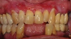
Figure 1 shows ulcerations and inflammation of the gingiva as well as sloughing on a recent visit before beginning treatment again with topical corticosteroids. Note the ulcerative areas especially superior to the canines.
Figure 2 depicts the same patient several weeks after treatment. The canine ulcerations are significantly improved.
The skin is fragile, and blisters producing vesicles and bullae are common. The lesions heal – but with scarring – and are often painful, requiring pain medication. Establishing a diagnosis can be difficult, and special immunoelectron microscopy evaluation of biopsied tissue is required if the disease is suspected.
Treatment most often consists of the use of topical and systemic corticosteroids. Immunosuppressive agents such as methotrexate, azathioprine, or cyclophosphamide may be used in some cases. In the case presented, dapsone cleared the skin lesions, but not the oral lesions. Ella is able to control the oral lesions with various topical corticosteroids. She has periods of spontaneous remission for a year or two but then has recurrence.
Due to the increasing age of the world population, autoimmune and blistering diseases may cause major problems in the elderly population, and we may continue to see more of the autoimmune blistering types of dieases as well as those combined with other diseases. Additionally, the amount of medications used by the older population continues to increase making the possibility of "medication or drug induced" disease states a reality as well.
Keep asking good questions, and always listen to your patients!
References
DeLong L, Burkhart NW. General and Oral Pathology for the Dental Hygienist. Lippincott Williams & Wilkins. Baltimore. 2008.
Ishii N, Hamada T, Dainichi T, Karashima T, Nakama T, Yasumoto S, Zillikens D, Hashimoto T. Epidermolysis bullosa acquisita: what's new? J Dermatol. 2010; 37:3:220-30.
Parker SR, MacKelfresh J. Autoimmune blistering diseases in the elderly. Clinics in Dermatology 2011;Feb. 29:1 69-79.
Plemons JM, Gonzales TS, Burkhart NW. Vesiculobullous diseases of the oral cavity. In: Disorders affecting the periodontium. Periodontology 1999;21:158-175.
Wright JT. Oral Manifestations of Epidermolysis Bullosa. Dermatol Clin 2010 Jan:28(1):159-64. PMID: 19945630.
Nancy W. Burkhart, BSDH, EdD, is an adjunct associate professor in the department of periodontics, Baylor College of Dentistry and the Texas A & M Health Science Center, Dallas. Dr. Burkhart is founder and co-host of the International Oral Lichen Planus Support Group (http://bcdwp.web.tamhsc.edu/iolpdallas/) and co-author of General and Oral Pathology for the Dental Hygienist. Her website for seminars is www.nancywburkhart.com.
Past RDH Issues


