TMJ assessment
How subjective is the process?
by Constance Schuster, RDH, BSExtensive forces can wreak havoc on the oral cavity. What about the temporomandibular joint and the constant movement it endures? Stress from excessive clenching and grinding or occlusal interferences may take a toll on this joint. The forces of the soft tissues must also be considered when evaluating intra- and extraoral changes. Dental professionals have the burden of determining dysfunction based on the absence or presence of signs and symptoms.
In reality, this evaluation is subjective in nature, because it is based on opinion or interpretation of information vs. an objective fact, which is verifiable based on concrete data.
Typical diagnostic tools in regard to movement would include radiographs, diagnostic casts, and the use of devices such as rulers and periodontal probes to record actual measurements. But when it comes to determining temporomandibular joint function, the evaluation is subjective due to the variation of the degree of signs or symptoms and the provider assessing the patient,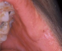
The temporomandibular joint is evaluated through palpation by placing the fingers over the joint while the patient opens and closes their mouth (see Figures 1, 2). The posterior portion of the condyle may be better assessed by placement of the little fingers into the patient's ears during open and close (see Figure 3). This evaluation is subjective, relying on the clinician's tactile sensitivity, pressure exhibited during palpation, and whether or not the clinician feels these signs.
Signs and symptoms evaluated during a TMJ analysis would include:
- Assessment for presence of crepitation and subluxation through palpation and listening with stethoscopy
- Presence of discomfort
- Limited opening
- Deviation of the TMJ upon opening or closure
We tend to forget the importance of the facial muscles and how necessary they are to mastication, speech, and our appearance. The muscles of the face need to work equally to help the mandible's gliding hinged joint function correctly. The temporalis, masseter and pterygoids are the muscles affected by the TMJ. The masseter, temporalis, and medial pterygoid help to elevate the mandible. To evaluate these muscles and determine their involvement in possible temporomandibular dysfunction, the patient should clench and the clinician palpate for signs of hypertrophy and discomfort.
The lateral pterygoid helps to protrude the mandible and creates lateral movement with unilateral contraction. This muscle will be extremely sensitive if TMD is present. Palpation of this muscle is performed intraorally while the mouth is partially closed. Pressure applied distal and buccal to the maxillary tuberosity digitally will evaluate this muscle's parafunctional stress.1
Clinical studies have demonstrated that palpation alone is less sensitive in detection of crepitation than the use of stethoscopy.2 Joint sounds can vary from popping, clicking, or grinding. At times it may be difficult to distinguish between these sounds because it could be a combination of a pop click or a grinding click. This assessment is up to the clinician to differentiate and record. However, the reliability of classifying joint sounds while utilizing palpation and auscultation is less than 50% from one clinician to the next.3 The variation demonstrates the lack of reliability of subjective evaluations in which the perception of the sound is relying on the absence of human error.
Temporomandibular dysfunction usually is categorized as one of the following: muscle disorder, disc displacement, or arthritis.4 A frequently utilized medical diagnostic code for the TMJ is ICD-9-CD 524.64, which is defined as temporomandibular joint sounds on opening and/or closing of the jaw. However, ICD-9-CM Diagnosis Code 524.69 (temporomandibular joint disorders, other specified temporomandibular joint disorders) encompasses all of the aforementioned categories for TMD.
One way to objectively evaluate the TMJ while putting an actual measurable number to sounds created during opening and closing is through the use of JVA (Joint Vibration Analysis) – a biometric technology that is becoming more popular in diagnosis and measurement of treatment success. BioRESEARCH Associates Inc. is a Milwaukee, Wis., company that provides different biometric equipment for the diagnosis and treatment of TMD (see Figure 4). These systems use objective criteria for defining TMJ sounds through electroacoustical technology.5
The initial development of biometric equipment to evaluate the TMJ began with electrosonography. This technology uses a pressure membrane, which records vibration through the air. It was not sensitive enough to discriminate the different sounds of the TMJ during function due to picking up external sounds such as sounds within the dental practice. Next, the electrovibratography was developed with a pressure sensor buried in silicone, which made it more sensitive to small vibration and shielded it from environmental sounds. This technology was able to determine the difference between the sounds of fluid, disc, and bone – therefore, creating a device with more diagnostic ability.
Without some type of recordable measurement, the typical dental office's assessment of the temporomandibular joint is subjective. Most assessments include deviation upon opening/closing, and whether or not crepitus/subluxation is present.
In reality, how can one measure success of treatment? Would it be only by the elimination of crepitus/subluxation? Using objective measurements from a recorded JVA scan, success can be determined through a decrease in joint vibrations. The subjective decrease in environmental noise from the use of shielded sensors also aids in the accuracy of recording only joint vibrations and not room noises. Preliminary studies have reported that most success for acute TMD from disc displacement, arthritis, or myofacial pain involves combination therapy that includes active exercises, manual therapy, postural correction, and relaxation techniques.6
Many practitioners feel apprehensive about treating TMD because of the many factors involved in assessment, diagnosis, and treatment of this disorder. However, as providers of dental care, it is our responsibility to assess and record our findings to the best of our ability. The doctor should determine the course of treatment and whether or not to refer the patient to another provider who is more experienced in the treatment of TMD. Or, if the doctor does treat the patient, he or she should be providing the best standard of care to the patient. Ultimately, we need to obey our Hippocratic oath to do no harm, and sometimes doing nothing is harmful. By providing the care needed by patients who are suffering from TMD, whether it be utilizing more objective diagnostic equipment, fabricating splints, or recommending/providing therapeutic exercise or relaxation techniques, we cannot be classified as stagnant professionals. We need to ask ourselves whether or not we are comfortable with the services we provide or can we strive to perform better. Following this basic philosophy guides each of us to provide the highest quality of care to our patients. Ultimately, excellence should be a goal for all dedicated professionals.
Constance Schuster, RDH, BS, received her associate's in applied science dental hygiene from Milwaukee Area Technical College and a Bachelor of Science degree in Biomedical Science from Marquette University. She studied Orofacial Myology is Los Angeles, Calif. and completed her internship in Pacific Palisades, Calif. She is a past president of Wisconsin Dental Hygienists' Association. Currently, she is president of Lotus Dental Solutions, LLC, providing orofacial therapy to patients and consulting/staffing services to dental practices. She has experience in dental assisting, dental hygiene, and field operations management. She has worked not only in general dentistry, but also orthodontics, pedodontics, prosthodontics, and periodontics. Her passion for dentistry continues to grow by mentoring hygienists and networking with other dental professionals through ADHA membership and involvement with CareerFusion.
References
- Woodall I, Dafoe B, Young N, Weed-Fonner L, Yankell S. Comprehensive Dental Hygiene Care. Third Edition 1989:282.
- Wabeke K, Spruijt R, van der Zaag J. The Reliability of Clinical Methods for Recording Temporomandibular Joint Sounds. Journal of Dental Research 1994:73:1157.
- Dworkin SF, LeResche L, Derouen T. Reliability of Clinical Measurement and Temporomandibular Disorders. Clin J of Pain, 1988, Volume 4 Number 2; 89-99.
- Roda R, Bagan J, Fernandez J, Bazan S, Soriano Y. Review of Temporomandibular Joint Pathology. Part I: Classification, Epidemiology, and Risk Factors. Medicina Oral Patologia, Oral y Cirugia Bucal 2007;12:E292-8.
- Hwang I, Jung D, Lee J, Kang D. Evaluation of TMJ sound on the subject with TMJ disorder by Joint Vibration Analysis. Journal of Advanced Prosthontics 2009; 1:26-30.
- Medlicott M, Harris S. A Systematic Review of the Effectiveness of Exercise, Manual Therapy, Electrotherapy, Relaxation Training, and Biofeedback in the Management of Temporomandibular Disorder. Physical Therapy 2006;86:955-973.
Past RDH Issues