Waiting to exhale: Managing sleep apnea for the pediatric dental patient

By Susan Woodley, RDH, and Lee Somerville, RDH, MS
Have you ever listened to a grandparent, parent, or spouse snoring so loudly you couldn’t sleep? No matter how hard you tried to ignore it, it was all you could focus on. Then, suddenly, sweet silence! You breathed a sigh of relief because the snoring stopped!
However, the only thing that stopped was this person’s breathing! Then the silence was broken and the “sound show” resumed as this person began frantically filling his or her lungs with air. The process was repeated over and over throughout the night. How many of us have shared these “snoring” stories in a social setting to collective laughter? These “snoring” episodes are actually part of a disease process called sleep disordered breathing (SDB), and it’s no laughing matter. SDB covers a broad spectrum of sleep disorders with obstructive sleep apnea (OSA) being the most common. It is significant to note that OSA, cancer, and AIDS are considered the most dangerous and destructive diseases on earth!1
In the past, OSA was believed to be directly related to increased body fat; however, more recent studies confirm there are other causative factors. These include overly relaxed throat and neck muscles due to eating or drinking too closely to bedtime; nasal or pharyngeal/laryngeal blockage; and flabby throat and oral structures (e.g., a large uvula or a floppy soft palate).
Exactly what is sleep apnea (SA)? SA occurs when air flow is completely stopped, and it is diagnosed when there are five apneas (i.e., cessation of breathing during sleep) or 10 apnea-hypopnea (i.e., when airflow is reduced by one-half to two-thirds) episodes per hour of sleep.2 Apnea alone is not a problem except when it exceeds 10 seconds in duration. There are three types of sleep apnea: central, obstructive, and mixed. Central apnea occurs when there is no respiratory effort due to brain stem immaturity of the respiratory control. This is commonly seen in premature babies and newborns. OSA is caused by airway obstruction. In young children, the obstruction is most often due to enlarged tonsils and adenoids. With a fivefold increase in childhood obesity in Canada and the United States over the last 15 years, we see an increasing number of children with obesity-related SDB.3,4 Mixed sleep apnea combines features of both central and obstructive causes. The Canadian Sleep Society (css-scs.ca) and the American Sleep and Breathing Academy (americansleepandbreathingacademy.com) are valuable resources on this topic.

Given the added complexity of behavioral and developmental issues that often accompany a child with SDB, our mission is to heighten the awareness for the dental professional and in doing so, promote earlier intervention. As cotherapists, it is important for us to have a clear understanding of our role in interdisciplinary management and to highlight various methodologies of treatment to broaden our scope of knowledge.
Contrary to adult SDB, pediatric SDB or OSA often presents with behavioral symptoms that can have cascading effects in many areas of their life. These may include mood changes, misbehavior, and poor school performance. Often symptoms of SDB are confused with ADHD and treated with medications that include stimulants, nonstimulants, and antidepressants. Not every child with academic or behavioral issues will have SDB, but if a child snores loudly on a regular basis and is experiencing mood, behavior, or school performance issues, SDB should be considered.
The largest and most comprehensive study to examine the effects of SDB symptoms on behavior from six months to seven years of age was conducted in the United Kingdom. The study evaluated and followed more than 11,000 children for over six years. The combined effects of snoring, apnea, and mouth-breathing on the behavior of children enrolled in the Avon Longitudinal Study of Parents and Children in the United Kingdom were examined. Parents completed questionnaires about their children’s SDB symptoms at several intervals, from 6 to 69 months of age. At approximately ages four and seven years old, parents filled out the Strengths and Difficulties Questionnaire (SDQ). The SDQ rated the individual for inattention/hyperactivity; emotional symptoms (anxiety and depression); peer difficulties; behavior problems (aggressiveness and rule-breaking); prosocial behavior (sharing, helpfulness, etc.).
The study controlled for 15 possible confounding factors (e.g., socioeconomic status, maternal smoking during the first trimester of pregnancy, low birth weight). The study revealed children with SDB were from 40% to 100% more likely to develop neurobehavioral problems by age seven, compared to children without breathing problems. The largest increase was seen in hyperactivity. There were, however, significant increases across all five behavioral measures. Children whose SDB symptoms peaked early, at six or 18 months, were 40% and 50% more likely, respectively, to have behavioral problems at seven years of age compared with children who had normal breathing. Children with the worst behavioral problems had SDB symptoms that continued throughout the evaluation period and became most severe at 30 months.5
This shines a light on the unique opportunity for dental professionals to be on the front line of discovery for numerous oral-systemic diseases. Many of our patients visit the dental office more often than any other health-care provider. Given its severity and far-reaching effects, should we not consider screening for SDB on a routine basis? The pathway to discovery begins with a clear understanding of the physical signs and symptoms of pediatric SDB:
- Abnormal breathing during sleep
- Bruxism
- Frequent awakenings or restlessness
- Frequent nightmares
- Enuresis (bedwetting)
- Difficulty awakening
- Excessive daytime sleepiness
- Hyperactivity/behavior problems
- Daytime mouth breathing
- Poor or irregular sleep patterns
- Growth impairment
The screening process for the dental professional begins with general observation of the patient. Mouth breathing and adenoidal facies (i.e., dull expression with open mouth) should be noted. Hyponasal voice (i.e., a voice that sounds like the nose is pinched when speaking) is a clue to nasal obstruction; a muffled voice suggests adenotonsillar enlargement. Secondly, the lateral facial profile should be examined for retrognathia, micrognathia, or midfacial hypoplasia. These can all have an effect on the nasopharyngeal and oropharyngeal passages and are key diagnostic findings. The oral cavity should be observed for tongue and soft palate size and appearance. A large tongue along with or independent of a high/low-arched or elongated palate may reveal a predisposition to SDB.6
Note the patient’s head posture. Blocked nasal breathing and jaw joint inflammation both result in forward head posture. Persistent forward head posture causes muscle contraction and results 96% of the time in occipital and forehead headaches.7
Once you have established and documented clinical findings, asking a series of questions contributes greatly to the screening process for SDB:
- Does the patient snore?
- Have you heard the patient grinding his or her teeth?
- Does the patient appear restless and/or awaken frequently at night?
- Does the patient have frequent nightmares?
- Does the patient wet his or her bed?
- Is it difficult to waken the patient in the morning?
- Are you aware of excessive sleepiness during the day?
- Are there any hyperactive or behavioural issues you are aware of?
- Would you consider the patient to be a mouth breather?
Contacting the physician
In keeping with an interdisciplinary approach, it is vital to share any abnormal clinical findings with the child’s physician as soon as possible. Children are in such a rapid state of cognitive development that time is of the essence to effect change. According to Halbower et al., “any alterations of health and brain function associated with SDB could permanently alter a child’s social and economic potential, especially if the disorder is not recognized early in life or treated inadequately. It is imperative that healthcare workers actively seek out symptoms of SDB in patients and educate parents and teachers about the signs and symptoms of SDB to improve early detection and treatment for prevention of long-term morbidity.”8
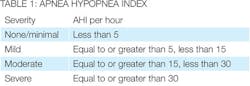
After a physical examination, the physician should prescribe a polysomnogram (a sleep study) to confirm the diagnosis of OSA in collaboration with the clinical suspicion, history, and physical findings from the dental professional. Once the polysomnogram is completed, severity is measured by the Apnea Hypopnea Index (AHI; see table 1).
A vast majority of cases of OSA in children are associated with adenotonsillar hypertrophy. The peak prevalence of childhood OSA is at two to eight years of age, which is the age when the tonsils and adenoids are the largest in relation to the underlying airway size. An endoscopy has shown the site of collapse is most often at the level of the adenoid.10
Although research indicates variances in the success rates from tonsillectomy and adenoidectomy, these procedures are often considered to be the first approach in management of pediatric SDB.11,12
Treatment options
Continuous positive airway pressure (CPAP) is another avenue in managing pediatric OSA. From a medical point of view, this treatment option is considered to be very successful when directions for use are followed carefully. Compliance does often present a challenge with inconsistencies in wear time, variances in caregiver philosophies, and other real-life situations that can impede the success rates associated with a CPAP.
Orthodontic appliances are gaining traction as a viable option in orthopedic management of SDB in the young patient. These appliances can be used as a prevention tactic as well as secondary therapeutic management with other modalities. The appliances used most often involve expansion of the palate or anterior posturing of the mandible and tongue during sleep. The significance of early detection and treating the underlying craniofacial abnormalities in children at high risk for developing OSA could, in effect, prevent the disorder. Since maxillary constriction is a feature of chronic nasorespiratory obstruction,13 rapid maxillary expansion has the potential to play a prominent role in a prevention strategy. The quality of sleep of these children improves after rapid maxillary expansion, regardless of the severity of their respiratory obstruction. Orthodontists who perform a comprehensive head-neck examination are uniquely positioned to identify children with malocclusion that could lead to development of SDB.14
Narrowing of the pharyngeal airway passage and adaptations in the soft palate are common among patients who present with a retrognathic mandible.15,16 As a result, the space between the cervical column and the mandible decreases, leading to a posteriorly postured tongue and soft palate. This increases the chances of impaired respiratory function during the day and possibly causing SDB symptoms, such as snoring, upper airway resistance syndrome, and OSA.17,18 A study was conducted to determine the effects of a twin-block appliance on the anatomy of the pharyngeal airway passage. Thirty-eight male and female subjects, age range of eight to 14 years, with skeletal class II malocclusion associated with mandibular retrusion participated in the study. It was concluded that correction of mandibular retrognathism by functional appliances improved the dimensions of the upper airway.
“Finding Connor Deegan” is a five-minute video revealing the “human” side of a child living with SDB. It illustrates the far-reaching effects SDB can have on an individual’s life and how that can all change with a proper diagnosis and management strategies. We encourage you to take a few minutes to watch this video on YouTube (search for “Finding Connor Deegan) and layer your learning experience.
Regardless of the treatment options, the individual with SDB is considered to be at high risk for dental caries and gingival inflammation.19 As dental professionals, we know scientifically that xerostomia plays a crucial part in the formation and adherence of plaque biofilm. Consistent mouth breathing and the use of functional appliances can contribute greatly to this undesirable environment. The Philips Sonicare For Kids Power Toothbrush, with 31,000 brush strokes per minute and high amplitude, creates patented fluid dynamic action to reduce the biofilm burden. With Bluetooth connectivity, the child can connect to an app that will help guide and coach his or her brushing experience in real time. Sonicare For Kids is safe and gentle for use with all appliances and removes 75% more plaque in hard-to-reach areas than a manual toothbrush in children aged seven to 10 years.20-22
The collaboration of an interdisciplinary team, clinical circumstances, research, and historical data all influence a customized approach to treating SDB in the young patient. As dental professionals, we want to prevent more to treat less. Implementing systematic screenings for SDB should be routine and the standard of care in every dental practice.
Evidence tells us that early interventions with pediatric SDB can have a profound effect in the formative years. We all know how it feels to save a tooth. Just imagine how great it would feel to change a life! RDH
Author’s note: This article was originally published in the November 2016 issue of OralHygiene magazine in Canada. Permission was granted to the authors for republication in RDH magazine. RDH
Lee Somerville, RDH, MS, is the manager of professional education in the northeast United States for Philips Oral Healthcare, and is based in Stamford, Conn. Susan Woodley, RDH, is the manager of professional education for Philips in western Canada.
References
1. Keropian B. Dentistry’s new frontier: the treatment of obstructive sleep apnea. http://www.dentistryiq.com/articles/2010/06/dentistry-s-new-frontier-the-treatment-of-obstructive-sleep-apnea.htm. Published June 2, 2010.
2. Parisi RA. Respiration and respiratory function: Technique of recording and evaluation. In: Chokroverty S, ed. Sleep Disorders Medicine: Basic Science, Technical Considerations, and Clinical Aspects. 2nd ed. Boston: Butterworth-Heinemann; 1999:220-221.
3. Anders TF, Eiben LA. Pediatric sleep disorders: a review of the past 10 years. J Am Acad Child Adolesc Psych. 1997;36(1):9-20
4. Katz SL, MacLean J, Hoey L, et al. Quality of life improvement with positive airway pressure therapy for sleep disordered breathing in obese youth. Am J Respir Crit Care Med. 2016;193:A1222. http://www.atsjournals.org/doi/abs/10.1164/ajrccm-conference.2016.193.1_MeetingAbstracts.A1222. 5. Bonuck K, Freeman K, Chervin RD, Xu L. Sleep-disordered breathing in a population-based cohort: Behavioral outcomes at 4 and 7 years. Pediatrics. 2012;129(4):e857-e865. doi: 10.1542/peds.2011-1402.
6. Verma SK, Maheshwari S, Sharma NK, Prabhat KC. Role of oral health professional in pediatric obstructive sleep apnea. Natl J Maxillofac Surg. 2010;1(1):35-40. doi: 10.4103/0975-5950.69162: PMCID: PMC3304178.
7. Simmons HC, Gibbs SJ. Anterior repositioning appliance therapy for TMJ disorders: specific symptoms relived and relationship to disc status on MRI. J Cranio Pr. 2005;23(2):89-99.
8. Halbower AC, McGinley BM, Smith PL. Treatment alternatives for sleep-disordered breathing in the pediatric population. Curr Opin Pulm Med. 2008 Nov;14(6):551-8.
9. Harvard Medical School. Understanding the results. Apnea: Understanding and Treating Obstructive Sleep Apnea website. http://healthysleep.med.harvard.edu/sleep-apnea/diagnosing-osa/understanding-results. Updated February 11, 2011.
10. Isono S, Shimada A, Utsugi M, Konno A, Nishino T. Comparison of static mechanical properties of the passive pharynx between normal children and children with sleep-disordered breathing. Am J Respir Crit Care Med. 1998;157(4 Pt 1):1204-12.
11. Friedman M, Wilson M, Lin HC, Chang HW. Updated systematic review of tonsillectomy and adenoidectomy for treatment of pediatric obstructive sleep apnea/hypopnea syndrome. Presented at: The Annual Meeting of the American Academy of Otolaryngology-Head and Neck Surgery; September 21-24, 2008; Chicago, IL. http://www.sciencedirect.com/science/article/pii/S0194599809000850.
12. Ahn YM. Treatment of obstructive sleep apnea in children. Korean J Ped. 2010;53(10):872-879. doi:10.3345/kjp.2010.53.10.872.
13. Jureyda S, Shucard DW. Obstructive sleep apnea - an overview of the disorder and its consequences. Semin Orthod. 2004;10(1):63-72.
14. Ashok N, Varma NK, Ajith VV, Gopinath S. Effect of rapid maxillary expansion on sleep characteristics in children. Contemp Clin Dent. 2014;5(4):489-94.
15. Kirjavainen M, Kirjavainen T. Upper airway dimensions in class II malocclusion. Effects of headgear treatment. Angle Orthod. 2007;77(6):1046-1053.
16. Jena AK, Singh SP, Utreja AK. Sagittal mandibular development effects on the dimensions of the awake pharyngeal airway passage. Angle Orthod. 2010;80(6):1061-7.
17. Schafer ME. Upper airway obstruction and sleep disorders in children with craniofacial anomalies. Clin Plast Surg. 1982;9(4):555-567.
18. Ozbek MM, Miyamoto K, Lowe AA, Fleetham JA. Natural head posture, upper airway morphology and obstructive sleep apnoea severity in adults. Eur J Orthod. 1998;20(2):133-43.
19. http://jcodental-uobaghdad-edu.org/index.php/jbcd/article/viewFile/216/pdf_105
20. Milleman J, Putt M, Olson M, Master A, Jenkins W, Schmitt P, Strate J. Comparison of plaque removal by Sonicare For Kids and a manual toothbrush in children aged 7-10 years. Int J Ped Dent. 2009;19:s1.
21. Defenbaugh J, Schmitt P, Master A, Jenkins W, Strate J. Brushing duration and use interaction patterns of manual versus sonic toothbrushes in children aged 7-10 years. Int J Ped Dent. 2009;19:s1.
22. Jenkins W, Master A, Defenbaugh J, Wei J; Philips Oral Healthcare. An observational in-home use test of children 4-10 years using Sonicare For Kids. J Dent Res. 2010;89:(spec iss B):Abstract 3696.