Insertion site: Height of the mucobuccal fold distal to the zygomatic process and superior to apex of the maxillary second molar. Angulations: Outward laterally at 45-degree angle from the midsagittal plane and downward 45-degree angle from the occlusal plane.
Dental hygienists routinely and comprehensively debride the challenging anatomy of the maxillary molar teeth. Anesthesia is frequently required especially for initial debridement procedures.
In fact, the maxillary molars are often the first to develop periodontal problems.1 The PSA block is preferred by this author when anesthetizing the maxillary molars because it reliably demonstrates a high success rate (greater than 95%)2 for profound and sustained anesthesia to the molar pulps and buccal periodontal and soft tissues with one injection, and because it is comfortable for the patient.
The alternative, supraperiosteal injections in the area, often requires multiple needle penetrations (three vs. one), more volume of anesthetic (1.8 mL vs. 0.9-1.8 mL), and usually provides a shorter duration and less profound anesthesia. When the PSA block is combined with the anterior superior alveolar (ASA) block using an infraorbital approach, often referred to as the infraorbital (IO) block, the entire maxillary arch on one side may be anesthetized (except palatal tissues).
When the PSA is combined with the anterior middle superior alveolar (AMSA) block, the entire hemimaxilla may be anesthetized (including palatal tissues).3 It is no wonder that dental hygiene programs require clinical competency for the PSA injection. According to a survey conducted by the author in November 2014, of the 95 responding dental hygiene education program directors (representing 39 states across the country), 95% required clinical competency for the PSA block injection.
Tips for Provision of PSA Block
1. 25-gauge short needle
2. Dry, topical, dry
3. Adequate retraction
4. Syringe barrel:
Outward 45 degrees from midsaggital plane
Downward 45 degrees from occlusal plane
5. Rapid penetration
6. Advance through soft tissue
16mm (normal adult skull)
7. Aspirate in three planes prior to deposition
8. Slow deposition
.09-1.8mL/60sec minimum
Respirate in same planeafter every 1/3 cartidge is dispensed
Technique
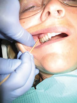
An important approach for providing the PSA block includes the insertion at the height of the mucobuccal fold distal to the zygomatic process and superior to the apex of the maxillary second molar and maintaining orientation of the syringe barrel outward laterally at a 45-degree angle away from the midsagittal plane and downward at a 45-degree angle away from the occlusal plane of the maxillary teeth4 (see Figure 1).
After assembling the proper armamentarium, prepare the patient’s tissues by drying the area of insertion, applying topical as directed, and drying the tissue again. The patient should be asked to partially open and slide the mandible toward the side of the injection. Retract the tissues both upward and outward to facilitate ease of injection. Adequate retraction is very important during the provision of the PSA so that both the angulation and depth of the needle may be observed. A 2X2 gauze may be helpful in keeping tissues dry so that adequate retraction will be maintained throughout the injection.
Resting the barrel of the syringe against the finger providing retraction can also provide additional stability for the syringe during advancement (see Figure 1). Establish a stable and safe fulcrum (extraoral or clinician’s body). Orient the 25-gauge SHORT needle (a 27 short may be used, but is not preferred) and the syringe barrel outward laterally at a 45-degree angle away from the midsagittal plane and downward at a 45-degree angle away from the occlusal plane of the maxillary teeth.
Insert the needle at the height of the mucobuccal fold distal to the zygomatic process and superior to the apex of the maxillary second molar. Advance the needle slowly, toward the target area—the PSA nerve as it enters the maxilla through the PSA foramina on infratemporal surface of the maxilla.2,5
Avoid depositing anesthetic solution before reaching the target. Advancement should not be uncomfortable for the patient as the needle should only be penetrating through soft tissues. Bone should not be contacted. If bone is contacted, the angle of the needle toward the midline is too great, and the needle should be withdrawn slightly and the barrel of the syringe brought closer to the occlusal plane and then re-advanced.2,5
For an adult skull, advance the needle 16mm (3/4 of short needle) and begin aspirations in 3 planes:
Aspirate at target
If negative, rotate syringe a quarter turn toward clinician and aspirate again
Finally, if negative, rotate syringe back to original position and aspirate again.5
After the consecutive negative aspirations, slowly deposit 0.8mL to 1.8mL (1/2-1 cartridge) of anesthetic over a minimum of 60 seconds. In her textbook, Logothetis recommends reaspirating, during deposition, in the same plane, after every ¼ cartridge is deposited.5
Special Circumstances
For approximately 28% of patients, the mesiobuccal root of the maxillary first molar is innervated by the middle superior alveolar (MSA) nerve instead of the PSA nerve.2 For these patients, a second injection, the MSA block, is required in order to innervate the mesiobuccal root of the first molar.
If anesthesia of the posterior palate is needed, the greater palatine (GP) block should be provided in addition to the PSA block. Often the palatal roots of the molars are also innervated by branches of the GP nerve, and provision of the GP block may be indicated to fully anesthetize those roots, particularly when there is furcation and root concavity involvement.5
When local anesthetic is deposited lateral to the target, some mandibular anesthesia may occur because the mandibular branches of the trigeminal nerve are located lateral to the PSA nerve. Patients may report some numbness of the tongue and/or lower lip.2,5
Common technique errors include the failure to maintain retraction and to maintain the 45-degree angle away from the midsagittal plane. Often, clinicians begin with proper technique, but subsequently relax retraction and close the angle to the midsagittal plane (allow syringe to move toward buccal aspect of teeth). This usually results in needle movement away from the target and/or insufficient advancement due to obscuring the view of the penetration site.
Some clinicians report that they avoid providing the PSA block due to the risk of hematoma (3%). Although the risk is not as high as with the IA or mental/incisive blocks which are 10-15% and 5.7% respectively,2 the hematoma which can result from the PSA block is the only hematoma that is visible extraorally.6 The risk of hematoma from a PSA injection can be minimized by using a short (to avoid over-insertion) 25-gauge needle, aspirating in 3 planes multiple times (to facilitate reliable aspiration) before and during the slow deposition of anesthetic.2,5-8
The PSA block is an important injection for dental hygienists to master and employ during pain control for a variety of reasons. It is safe, extremely reliable, and very effective when care is taken to follow proper technique. Since dental hygienists often treat wide areas of the mouth during one appointment, limiting both the number of penetrations as well as the volume of anesthetic by using nerve blocks, such as the PSA is prudent. The PSA block when combined with other injections which anesthetize wide areas, such as the AMSA and ASA blocks, promotes efficiency as well as safety and comfort for the patient.

LAURA J.WEBB, RDH, MS, CDA, is an experienced clinician, educator, and speaker who founded LJW Education Services (ljweduserv.com). She provides educational methodology courses and accreditation consulting services for allied dental education programs and CE courses for clinicians. Laura frequently speaks on the topics of local anesthesia and nonsurgical periodontal instrumentation. She was the recipient of the 2012 ADHA Alfred C. Fones Award. Laura can be contacted at [email protected].
References
1. Beemsterboer P. 2014 Periodontology for the Dental Hygienist, 4th ed; Elsevier.
2. Malamed S. 2013 Handbook of Local Anesthesia 6th ed; Elsevier.
3. Webb, L. The AMSA nerve block: Pair with the PSA nerve block for hemimaxillary anesthesia. RDH magazine.Aug 2017 Vol 37 No 8
4. Bassett, DiMarco, Naughton. 2010 Local Anesthesia for Dental Professionals; Pearson.
5. Logothetis D. 2017 Local Anesthesia for the Dental Hygienist, 2nd ed; Elsevier.
6. Malamed, S. Complications in local anesthesia administration. Dimensions of Dental Hygiene. Oct 2006. 4(10): 28-33
7. Blanton, Jeske. Avoiding complications in local anesthesia induction - Anatomical considerations. J Am Dent Assoc. 2003. 134 (7): 888-893.
8. Freuen ND, Feil BA, Norton NS. The clinical anatomy of complications observed in a posterior superior alveolar nerve block.The FASEB Journal 2007. 21:776-94.
About the Author

Laura J. Webb, MS, RDH, FAADH
Laura J. Webb, MS, RDH, FAADH, is an experienced clinician, educator, and speaker who founded LJW Education Services (ljweduserv.com). She has provided educational methodology courses and accreditation consulting services for allied dental education programs and CE courses for clinicians. Laura has frequently spoken on the topics of local anesthesia and nonsurgical periodontal instrumentation. She was the recipient of the 2012 ADHA Alfred C. Fones Award. Laura can be reached at [email protected].
Updated May 2023