BY NANCY W. BURKHART, BSDH, EdD
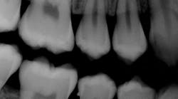
The patient today is a 15-year-old female who has a scheduled appointment for a dental prophylaxis with radiographs. While viewing the radiographs, the hygienist notices the presence of an enamel pearl on No. 29 distal (see Figure 1). The patient did not bring any previous radiographs with her for the appointment and, fortunately, radiographs were taken before the scaling procedure was performed. If scaling had been completed prior to recognizing that the enamel pearl was present, the appointment may have taken a totally different course. The enamel pearl would have been discovered during scaling; the potential for a broken instrument tip lodging in the tissue would have been a possibility.
So what is the etiology of the enamel pearl, and what is the significance for a dental professional?
Etiology: The enamel pearl is considered an abnormality. It is a developmental, localized formation of enamel occurring on the root surface of a tooth. It is a remnant of Hertwig's epithelial root sheath in which an area of stellate reticulum (the middle layer of the enamel organ) is retained between the two layers of epithelium that normally comprise the root sheath (Kessler, 2013).
According to Kessler: "The presence of the stellate reticulum allows differentiation of the root sheath cells into ameloblasts, leading to the formation of a small area of enamel on the external root surface."
In addition, literature also reports, "Enamel pearls may consist entirely of enamel connected to cementum or root dentin or may show incorporation of a cone of dentin without pulpal extension" (Sharma et al. 2013).
There is also a differentiation of the enamel pearl and the enamel projection. The enamel projection may extend into long, narrow projections and sometimes connect the enamel pearl to the root or crown portion (Risnes et al. 2000). Versiani et al. (2013) note other names that have been synonymous with the enamel pearl: enamel droplet, enamel nodule, enamel globule, enamel knot, enamel exostose, enameloma, and adamantoma.
Clinical features: The enamel pearl typically appears as a round, single, solid formation on the tooth root surface. The enamel pearl may vary in size from microscopic to a few millimeters. A dentist or hygienist may be cognizant of the nodule during a scaling procedure or as the pearls are detected in viewing the patient's radiographs before initiating a scaling procedure.
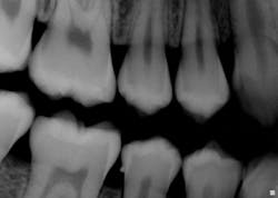
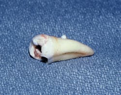
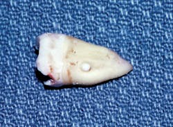
Radiographs/distinguishing characteristics: Radiographically, the enamel pearl is observed as a smooth, radiopaque structure on the root of a multirooted tooth. Since the nodule is below the tissue, it is not noted during the usual oral tissue exam. Enamel pearls are comprised of combinations of pure enamel and sometimes dentin. The dentin core may be found within the nodular entity and it is not uncommon to also detect a pulp horn as well. Pulp vitality testing is needed (see Figures 2, 3).
Epidemiology: The enamel pearl is most often found in the furcation areas of molar teeth and at the cemento-enamel junction. Maxillary teeth are most often affected and they occur more often on the buccal or lingual areas of mandibular teeth. Asians tend to be affected most often. A retrospective study by Colak et al. (2014) evaluated radiographs from 6,912 Turkish patients, out of which 5.1% of the study group exhibited enamel pearls. The researchers found a higher rate in males, 6.58% compared to females at 3.96%. The researchers in this study found a higher rate of enamel pearls in the mandibular first molar region with no statistical differences noted in the right or left side.
Differential diagnosis: Differential diagnosis considerations in confirming the enamel pearl may include:
• Calculus
• Enamel projections
• Pulp stones
• A composite restoration
Because the enamel pearl often results in deep pocket formation over time, the practitioner may assume that the structure is calculus. It would be assumed that the calculus was the cause of the deeper pocket and inflammation. It is also believed that the tissue areas adjacent to the enamel pearl have a weaker attachment to the periodontal ligament and these may be at higher risk for periodontal involvement and inflammation.
Treatment and prognosis: Early diagnosis of the enamel pearl is important since tissue destruction, development of periodontal pockets, and inflammation are known to occur with the entity on the root surfaces. Bacterial accumulation around the structure may result in tissue destruction.
Figure 1: Courtesy of Dr. Kathryn Savitsky, Charlotte, N.C.
Figure 2: Courtesy of Dr. Harvey P. Kessler, Baylor College of Dentistry, Texas A & M Health Science Center, Dallas, Texas.
Figure 3: Courtesy of Dr. Harvey P. Kessler, Baylor College of Dentistry, Texas A & M Health Science Center, Dallas, Texas.
Virsiani (2013) describes this process related to the enamel pearl as promoting enhanced plaque retention and protecting oral microorganisms from the action of salivary enzymes and oral hygiene measures. This may predispose a particular site to periodontal problems.
Kessler (2013) points out that the structure is solid and repeated attempts to dislodge the enamel pearl may result in a broken instrument tip. Additionally, because some enamel pearls may contain pulp horns, attempted removal with an instrument may necessitate endodontic treatment. When the enamel pearl is in the furcation areas or extending toward the posterior cervical root area, surgical procedures are often necessary to remove the structure.
As always, thoroughly observe and listen to your patients, and always ask good questions! RDH
References
Colak H, Hamidi MM, Uzgur R, Ercan E, Turkal M. Radiographic evaluation of the prevalence of enamel pearls in a sample adult dental population. Eur Rev Med Pharmacol Sci. 2014; 18:440-444.
Kessler HP. Chapter 21: Pathology of Teeth. Delong L, Burkhart NW, Editors, 2nd ed. General and Oral Pathology for the Dental Hygienist. Wolters Kluwer Health, Lippincott Williams & Wilkins-Baltimore. 2013.
Sharma S, Malhotra S, Baliga V, Hans M. Enamel pearl on an unusual location associated with localized periodontal disease: A clinical report. J Indian Soc Periodontol. 2013 Nov-Dec; 17(6): 796-800.
Risnes S, Segura JJ, Casado A, Jimenez-Rubio A. Enamel pearls and cervical enamel projections on 2 maxillary molars with localized periodontal disease. Case report and histological study. Oral Surg Oral Med Oral Pathol Oral Radiol Endod 2000;89:493-7.
Versiani MA, Cristescu RC, Saquy PC, Pécora JD, de Sousa-Neto MD.
Enamel pearls in permanent dentition: case report and micro-CT evaluation. Dentomaxillofac Radiol 2013; 42: 20120332.
NANCY W. BURKHART, BSDH, EdD, is an adjunct associate professor in the department of periodontics, Baylor College of Dentistry and the Texas A & M Health Science Center, Dallas. Dr. Burkhart is founder and cohost of the International Oral Lichen Planus Support Group (http://bcdwp.web.tamhsc.edu/iolpdallas/) and coauthor of General and Oral Pathology for the Dental Hygienist. She was a 2006 Crest/ADHA award winner. She is a 2012 Mentor of Distinction through Philips Oral Healthcare and PennWell Corp. Her website for seminars on mucosal diseases, oral cancer, and oral pathology topics is www.nancywburkhart.com.


