The oral cavity fosters a complex interplay between diverse viral agents and host tissues. Human papillomavirus (HPV) and herpes simplex virus (HSV) emerge as key players, orchestrating a spectrum of distinct clinical presentations within the oral cavity.
Human papillomavirus
For decades, HPV was primarily recognized as the causative agent of cervical cancer and other anogenital malignancies. However, in recent years, there has been a paradigm shift: a growing body of evidence links specific HPV types to diverse clinical manifestations within the oral cavity.
HPV is a double-stranded DNA virus that comprises more than 200 subtypes, with some exhibiting tropism for mucosal and cutaneous tissues. In the oral context, specific HPV types are associated with distinct clinical sequelae.1 Studies in North America and China revealed a prevalence of HPV DNA in oral samples of healthy populations that ranged from 2%–15%.2 High-risk HPV types are linked to about 5% of all cancers globally, totaling 600,000 cases each year.3
More about HPV ... Beyond traditional screening: Incorporating oral HPV testing
Transmission primarily occurs through close oral-mucosal contact, including kissing, oral sex, and shared utensils. Notably, while some oral HPV infections resolve spontaneously, certain high-risk types can persist and contribute to the development of precancerous and cancerous lesions.
Based on their potential to cause disease, they are classified according to their risk level4:
- High-risk HPV is associated with an increased risk of developing cancer, particularly cervical and head and neck cancer. Examples include HPV-16, HPV-18, HPV-31, HPV-33, HPV-45, HPV-52, and HPV-58.5
- Low-risk HPV is generally not associated with cancer but can cause benign lesions such as common warts and genital warts. Examples include HPV-6 and HPV-11.
- Probable high-risk HPV types have limited data but show possible links to cancer development, needing further research for definitive classification. Examples include HPV-26, HPV-53, and HPV-66.
More than just warts: Differentiating common oral HPV manifestations
There is a surprisingly diverse range of clinical manifestations of HPV in the oral cavity. From harmless bumps to potentially precancerous lesions, understanding these different presentations is crucial for accurate diagnosis and timely intervention.
Focal epithelial hyperplasia (FEH)
FEH, or Heck’s disease, is a benign condition affecting the oral mucosa. It is commonly linked to HPV-13 and HPV-33.6 Moreover, it presents as asymptomatic, small, white, or pink papular lesions often found on the dorsal aspect of the tongue, lips, palate, or gingiva that typically self-resolves. Still, persistent lesions warrant consideration for biopsy to rule out dysplasia/carcinoma.7
Verruca vulgaris
Common warts are caused by various HPV types (e.g., HPV-2, HPV-4, HPV-27, HPV-57). They typically appear on the hands, feet, and soles, causing few to no symptoms. Verruca vulgaris is most commonly found on the skin. These warts can also occur in the mouth (but rarely), and when they do, they present as rough, elevated, white, or gray papules, most frequently on lips, gingiva, and palate. While they can occur at any age and are similar in their prevalence between the genders, they are most often diagnosed in individuals between their 30s and 50s. Due to its differential diagnosis of focal epithelial hyperplasia, condyloma acuminatum, oral squamous papilloma, oral lichen planus, oral leukoplakia, oral verrucous carcinoma (VC), and squamous cell carcinoma, a biopsy is necessary.8
Condyloma acuminatum
Condyloma acuminatum is caused by high-risk HPV types (HPV-6 and HPV-11). It primarily affects anogenital tissues, but oral transmission can occur. This type requires a swift diagnosis and management to prevent further transmission. Clinically, the lesions look like sharply pointed, cauliflower-like papillomatous growths that can be small, painless, white, pink, or flesh-toned bumps. These bumps may be single or appear in clusters, and they might be located on the tongue, lips, gums, or buccal mucosa.9
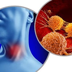
Oropharyngeal squamous cell carcinoma (OPSCC)
OPSCC affects the tonsils, the base of the tongue, and the soft palate. Its appearance is particularly alarming as it occurs despite declining smoking rates and is a known risk factor for head and neck cancers. This shift points to a new culprit: HPV, especially high-risk types HPV-16 and HPV-18 (with HPV-16 dominating 90% of the cases). Notably, this link is strongest in nonsmokers, unveiling a previously unrecognized dimension of OPSCC development, demanding further investigation into the role of HPV.10
Why verrucous carcinoma isn’t always linked to HPV
Don’t judge a wart by its looks. All the lesions mentioned above are part of the verrucous lesions group, characterized by their rough, wartlike appearance. Although some are mere bumps on the road, others raise concerns due to their potential link to malignancy. Therefore, including VC in this group is natural. Although the relationship between VC and HPV is complex and not fully understood (there is limited evidence of the association between VC and HPV), studies have yielded conflicting results, with some showing HPV DNA (particularly low-risk types like HPV-6 and HPV-11) in a minority of VC cases, while others find no association even when HPV is detected. HPV’s role in VC development is unclear.
Recognizing the diverse presentation of HPV lesions, understanding their potential causes, and employing appropriate diagnostic measures are crucial for accurate diagnosis and timely intervention. Early detection and proper management, particularly for potentially precancerous and malignant lesions, ensure optimal patient outcomes and safeguard oral health.
Understanding oral herpes: From tingles to blisters
HSV infection is a major global health concern, especially when it comes to orofacial manifestations. While commonly known for causing the characteristic blistering of “cold sores,” the clinical spectrum of oral HSV is much wider. HSV is a double-stranded DNA virus that comes in two subtypes: HSV-1 and HSV-2.
A familiar foe: Oral manifestations of HSV-1
Approximately 67% of adults in the US carry a ubiquitous, yet unwelcome, passenger: HSV type 1 (HSV-1). While asymptomatic in most individuals, HSV-1 can manifest as the well-recognized “cold sore,” also known as herpes labialis.
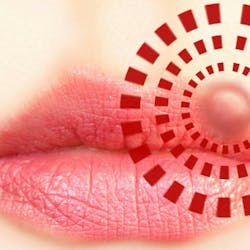
The initial viral invasion occurs through oral mucosa or skin breaches, allowing the virus to establish latency within the trigeminal sensory ganglia. Reactivation, triggered by diverse factors such as stress or UV radiation, leads to the characteristic clinical picture. Its prodromal phase features a tingling or burning sensation at the affected site, often the lip. This feeling will announce the eruption of small, erythematous vesicles filled with clear fluid. Progression involves vesicle rupture, crusting, and resolution within seven to 10 days.

Diagnosis typically relies on its clinical presentation, with viral culture or polymerase chain reaction (PCR) assays employed for atypical cases or patient reassurance. However, the scope of oral manifestations extends beyond classic cold sores. HSV-1 can present as herpetic gingivostomatitis, particularly in children, manifesting as painful oral ulcerations and vesicles.11 Moreover, atypical presentations involving the tongue, palate, or buccal mucosa can pose diagnostic challenges.
Accurately diagnosing and managing oral HSV-1 requires a nuanced understanding of its diverse clinical spectrum. This aids differentiation from other vesiculobullous conditions and ensures optimal antiviral therapy.
HSV-2 and the oropharyngeal landscape
While HSV-2 is primarily associated with genital herpes, its presence in the oropharyngeal region holds significant implications. Less common than HSV-1, oral HSV-2 infections can occur through oral-genital contact, presenting unique challenges in diagnosis, management, and transmission control.
The clinical presentation of oral HSV-2 often mirrors its genital counterpart, featuring painful vesicular lesions that progress to ulceration. However, primary infection with HSV-2 in the oropharynx can be more severe, exhibiting extensive vesicular eruptions and pronounced lymphadenopathy. Unlike HSV-1, oral HSV-2 typically manifests on the soft palate, tonsils, and posterior tongue, less frequently involving the lips and perioral region.12
A critical aspect of oral HSV-2 is its propensity for asymptomatic shedding, even without visible lesions. This shedding poses a significant risk for transmission, making diagnosis using different diagnostic techniques, such as viral culture or PCR testing, crucial.
Treatment for oral HSV-2 primarily involves antiviral therapy to shorten the course of infection, reduce symptoms, and suppress viral shedding. However, no cure exists, and recurrent outbreaks remain a potential concern.
Dental professionals play a vital role
Management of HPV and HSV infections in the oral cavity depends on the specific diagnosis and clinical presentation. While antiviral medications can alleviate symptoms and shorten outbreak duration, no cure exists for either virus. Preventive measures should focus on vaccination. HPV vaccines protect against high-risk HPV types linked to OPSCC and some oral FEH types. By understanding our role, we must highlight the importance of regular dental checkups for early detection and management of oral HPV and HSV infections, promoting preventive measures, and providing supportive care throughout the management process.
Editor's note: This article appeared in the April/May 2024 print edition of RDH magazine. Dental hygienists in North America are eligible for a complimentary print subscription. Sign up here.
References
- Bravo IG, Félez-Sánchez M. Papillomaviruses: viral evolution, cancer and evolutionary medicine. Evol Med Public Health. 2015;2015(1):32-51. doi:10.1093/emph/eov003
- Kreimer AR, Bhatia RK, Messeguer AL, Gonzalez P, Herrero R, Giuliano AR. Oral human papillomavirus in healthy individuals: a systematic review of the literature. Sex Transm Dis. 2010;37(6):386-391. doi:10.1097/OLQ.0b013e3181c94a3b
- Testi D, Nardone M, Melone P, Cardelli P, Ottria L, Arcuri C. HPV and oral lesions: preventive possibilities, vaccines and early diagnosis of malignant lesions. Oral Implantol (Rome). 2016;8(2-3):45-51. doi:10.11138/orl/2015.8.2.045
- Syrjänen S. Oral manifestations of human papillomavirus infections. Eur J Oral Sci. 2018;126 Suppl 1(Suppl Suppl 1):49-66. doi:10.1111/eos.12538
- Johnson DE, Burtness B, Leemans CR, Lui VWY, Bauman JE, Grandis JR. Head and neck squamous cell carcinoma. Nat Rev Dis Primers. 2020;6(1):92. doi:10.1038/s41572-020-00224-3. Erratum in: Nat Rev Dis Primers. 2023;9(1):4. doi:10.1038/s41572-023-00418-5
- Bendtsen SK, Jakobsen KK, Carlander ALF, Grønhøj C, von Buchwald C. Focal epithelial hyperplasia. Viruses. 2021;13(8):1529. doi:10.3390/v13081529
- Prabhat MPV, Lakshmi CR, Madhavi NS, Bhavana SM, Sarat G, Ramamohan K. Multifocal epithelial hyperplasia of oral cavity expressing HPV 16 gene: a rare entity. Case Rep Dent. 2013;2013:871306. doi:10.1155/2013/871306
- Sen S, Sen S. Intraoral verruca vulgaris. J Ayub Med Coll Abbottabad. 2021;33(2):354.
- Piña AR, Fonseca FP, Pontes FSC, et al. Benign epithelial oral lesions – association with human papillomavirus. Med Oral Patol Oral Cir Bucal. 2019;24(3):e290-e295. doi:10.4317/medoral.22817
- Hübbers CU, Akgül B. HPV and cancer of the oral cavity. Virulence. 2015;6(3):244-248. doi:10.1080/21505594.2014.999570
- Huang CW, Hsieh CH, Lin MR, Huang YC. Clinical features of gingivostomatitis due to primary infection of herpes simplex virus in children. BMC Infect Dis. 2020;20(1):782. doi:10.1186/s12879-020-05509-2
- Rathbun MM, Szpara ML. A holistic perspective on herpes simplex virus (HSV) ecology and evolution. Adv Virus Res. 2021;110:27-57. doi:10.1016/bs.aivir.2021.05.001
Andreina Sucre, MSc, RDH, is an oral pathology and oral surgery specialist, and a practicing dental hygienist in South Florida. She’s a speaker, writer, and advocate for early pathological diagnosis. Andreina was born in Venezuela where she earned her dental degree in 2002 from Universidad Santa Maria. In December 2005, she earned her certificate and specialization degree in oral pathology and oral surgery from Pontificia Universidad Javeriana, in Colombia. Follow her on Instagram @ThePathoRDH.