A hygienist of 40 years shares her enthusiasm with dental technology — specifically surgical microscopes — with her colleagues
by Susan Tompkins, RDH
The practice of dental hygiene has certainly changed in the past 40 years since I graduated from Fones School of Dental Hygiene, University of Bridgeport in Bridgeport, Conn. When I graduated, hygienists still wore nursing caps. There were no gloves or eye protection! We were not permitted to scale "below the sulcus."
I was fortunate to be able to stay at home with our four children until 1983, and when I returned to practice I already was met with changes in my profession. It is an understatement to say that our profession has become more sophisticated! We no longer perform "prophies," but provide scaling, root planing, and soft-tissue management. In the past, we had to rely heavily on our tactile sense, but the surgical microscope allows us to actually see what we are doing!
I work in Charlottesville, Va., for Dr. Wayne Remington. Dr. Remington became interested in the surgical microscope for two reasons. One of his colleagues was forced to give up his dental practice due to severe back and neck problems. My employer attended a dental esthetics course given by Dr. Cherilyn Sheets, where he observed her treating patients with the aid of a surgical microscope. That's when he realized that he could practice excellent dentistry sitting in an upright position.
After practicing with the surgical microscope, Dr. Remington developed the Virginia Microscope Training Center in Charlottesville. His goal is to encourage doctors to utilize the surgical microscope for all of their procedures and to provide this training with the use of mannequins mounted to regular dental chairs with surgical microscopes vs. other laboratory settings with limited arm motion and tabletop microscopes. Dr. Remington's desire is that at the end of his weekend course, the attendee will be able to perform dentistry in all four quadrants of the mouth. He has taught courses not only in Charlottesville, but in Tucson and Phoenix, Ariz., as well as in Europe.
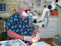
I have had the opportunity to complete Dr. Remington's training, as did my two colleagues. In addition, Dr. Remington has provided us with surgical microscopes in our operatories for the last two years. I remember the first day of the course and how excited I was to begin. Working through the microscope was nothing like I had anticipated. I felt like my hands, eyes, and brain were all disconnected! As a matter of fact, when evening came I was exhausted and went home directly to bed! But with Dr. Remington's guidance and encouragement, using the microscope became more natural. At this point, I cannot imagine working without it!
The microscope has the option to attach a digital camera or video recorder to it, which provides visual aids for the patient and documentation for the dentist. It is incredible to demonstrate to patients their need for specific treatment when they can see it for themselves.
Dental loupes have been a large part of my dental hygiene career. When they were first introduced to me, I thought they were the standard of practice. However, having adequate light with loupes is a challenge. Light intensity is determined by the inverse square law, which states that the amount of light received from a source is inversely proportional to the square of the distance.1 For instance, if a light source is two feet away from the patient and you move it only one foot away, the light will be four times as intense as it was at two feet. The surgical microscope has its own built-in light source, and with the light closer to the object, it is more intense. Also, the microscope offers three to six times the levels of magnification compared to dental loupes. This magnification enables the clinician to have a more relaxed eye position and reduces eye fatigue.
The dental chair that is used with the microscope is designed to keep the clinician upright, with back straight, neck straight, elbows resting on the chair arms, and feet flat on the floor. This position eliminates head, neck, and back fatigue.
The microscope assists me in the cursory diagnosis and assessment of each patient. I use it on both pediatric and adult patients. It has given me the opportunity to see areas of decay that may not have been diagnosed before and treat them before they progress. The patient feels confident that our clinical staff is providing the most precise dentistry and reflects a positive response due to the quality of their treatment.
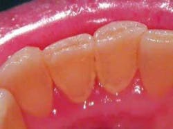
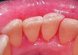
Here are examples of the digital capabilities of the surgical microscope capturing an image before and after treatment of a patient's lower central incisors. The images show a moderate to heavy accumulation of calculus that the microscope can magnify beyond that of the human eye or dental loupes.
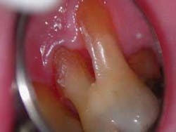
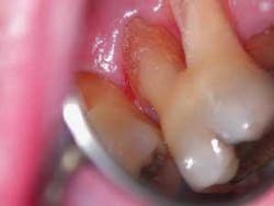
The following images indicate furcation calculus on the upper right first permanent molar. Again, the image is magnified beyond that of the human eye and dental loupes, allowing the clinician easier access.
So, I must tell you that being a "mature" hygienist, the impact of my new posture while treating eight patients a day is much healthier. I"m not bending over patients, craning my neck to see the distal of the upper second molars. I have learned to use my mirror to access difficult areas. I have found that the use of a narrower instrument cutting edge is required, because I can now see the instrument slide into the surface I want to scale. One of the companies that provides a thin blade scaler is American Eagle. These instruments are designed using XP technology, thus producing an instrument that requires no sharpening. Just recently, American Eagle introduced the Quick Tip system with a replaceable cone socket tip. The shanks of these scalers are relatively flexible, and the blades are very fine and sharp. EverEdge also has introduced a finer tipped curette called the Mini Five Gracies.2 Magnification and illumination are great assets when using ultrasonic scalers. They provide a clearer view in deep pocket areas and furcations.
I am able to more efficiently detect supragingival and subgingival calculus. In some cases, I'm able to see into the base of the pocket. The microscope assists in the detection of fractures, open margins, and accurate placement of sealants. Interestingly enough, patients (both adults and children) seem to stay in position in the headrest beneath the microscope light, which reduces the amount of time I spend repositioning the patient’s head.
My goal is to introduce this technology to the dental hygiene profession, because I know it will not be available unless it is provided by the dentist. Money is no longer an obstacle, because some entry-level microscopes cost approximately as much as an intraoral camera. Some companies provide leasing options for small monthly payments.
I encourage my fellow hygienists to investigate this new wave of dental treatment. It is an advantage for clinicians and an asset to patients. If you currently work for a dentist who uses a microscope, ask for training and a microscope of your own. It is an exciting time for the dental hygiene profession. We have equipment accessible to assist us in providing precision and excellence in treatment to our patients!
For more information, please visit http://www.microscopetraining.com. The author does not receive any compensation from companies or products mentioned in this article.
About the Author
Susan Tompkins, RDH, graduated from Fones School of Dental Hygiene, University of Bridgeport in Connecticut in 1967 with an associate’s degree. She has practiced for 26 years in Charlottesville, Virginia. She and her husband have four grown children and four grandchildren with one "on the way." Sue completed the surgical microscope training in 2005. She and her husband enjoy traveling.
References
1. Carr GB. Magnification and illumination in endodontics.
2. Pattison AM. Dimensions of Dental Hygiene.
