Case Study
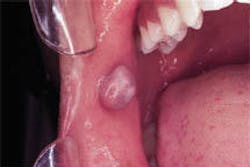
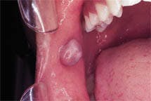
A 19-year-old female visited a general dentist for an initial examination. During the intraoral exam, a large, dome-shaped mass was noted on the buccal mucosa.
History
The patient was aware of the large, dome-shaped lesion on the inside of the cheek and stated that it had been present for at least several months, perhaps longer. The lesion was described as painless and had no history of bleeding. When questioned about trauma, the patient recalled biting her cheek repeatedly prior to the appearance of the lesion.
The patient appeared to be in a general good state of health and denied any history of serious illness. A review of the medical history revealed no significant findings. At the time of the dental appointment, the patient was not taking any medications.
Examination
The patient's blood pressure, pulse rate, and temperature were all found to be within normal limits. No enlarged lymph nodes in the head and neck region were detected upon palpation. Oral examination revealed one large, dome-shaped lesion on the anterior buccal mucosa (see photograph). Palpation of the lesion revealed a well-circumscribed, firm mass. Further examination of the oral tissues revealed no other lesions present.
Clinical diagnosis
Based on the clinical information presented, which of the following is the most likely clinical diagnosis?
- pleomorphic adenoma
- neurofibroma
- mucocele
- fibroma
- lipoma
Discussion
The fibroma (also known as irritation fibroma, traumatic fibroma, or focal fibrous hyperplasia) is a common oral lesion that arises secondary to chronic irritation or trauma. It is the most common swelling of the buccal mucosa and represents a response of connective tissue cells to chronic irritation. The fibroma is considered to be a reactive lesion. When traumatized, the tissues of the oral cavity react and an exuberant repair process is seen.
As a result, an overabundance of fibrous connective tissue is produced and the formation of a nodule or mass.
Clinical features
The fibroma is most often seen in patients between the ages of 30 and 50, and females are affected more frequently than males (2:1). There is no race predilection. The fibroma may occur in any area; the areas most frequently affected include the areas most easily traumatized, such as the buccal mucosa, tongue, and labial mucosa.
The typical fibroma appears as a sessile, dome-shaped mass with a smooth surface. The size of the lesion may range from 1-2 centimeters in diameter. The fibroma is usually pale pink in color, resulting from the increased amount of collagen in the lesion. Occasionally, the lesion may appear reddish. Most fibromas exhibit a smooth surface. If traumatized, ulceration on the surface may be noted.
The fibroma is firm upon palpation and exhibits a well-defined periphery. The fibroma is asymptomatic; no pain is associated with this lesion because nerve tissue does not proliferate along with the connective tissue produced in response to irritation.
Diagnosis and treatment
A diagnosis of a fibroma is based on its clinical appearance, history, and histologic examination. A history of trauma or chronic irritation to the area followed by the development of a sessile, firm mass is characteristic. Biopsy and histologic examination are necessary to establish a definitive diagnosis.
The recommended treatment for fibroma is surgical excision. In addition to surgical removal of the lesion, if the source of chronic irritation can be identified, it must be corrected as well. If the precipitating factor is removed, the lesion should not recur.
Joen Iannucci Haring, DDS, MS, is an associate professor of clinical dentistry, Section of Primary Care, The Ohio State University College of Dentistry.

