CASE STUDY
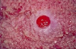
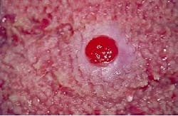
A 63-year-old male visited a general dentist for an initial examination. During the intraoral exam, a small red lesion was noted on the dorsal tongue.
History
The patient was aware of the small red lesion on the tongue and stated that it had been present for at least several months. The lesion was described as painless and had no history of bleeding. When questioned about trauma, the patient claimed that he often bites his tongue while chewing.
The patient appeared to be in a general good state of health and denied any history of serious illness. A review of the medical history revealed a hospitalization for a knee replacement. At the time of the dental appointment, the patient was not taking any medications.
Examination
The patient's blood pressure, pulse rate, and temperature were all found to be within normal limits. No enlarged lymph nodes in the head and neck region were detected upon palpation. Oral examination revealed a small, elevated red lesion on the dorsal tongue (see photo). Palpation of the lesion revealed a well-circumscribed and soft mass. Further examination of the oral tissues revealed no other lesions present.
Clinical diagnosis
Based on the clinical information presented, which of the following is the most likely clinical diagnosis?
- pleomorphic adenoma
- peripheral giant cell granuloma
- pyogenic granuloma
- granular cell tumor
- lipoma
Diagnosis
- pyogenic granuloma
Discussion
The pyogenic granuloma is a reactive overgrowth of tissue that occurs in response to trauma or local irritants. The term pyogenic granuloma is a misnomer, as the lesion is neither pus-producing as the word pyogenic suggests, nor a mass of granulation tissue, as the term granuloma implies. The pyogenic granuloma is a common lesion that may be found in the oral cavity as well as on the skin.
Clinical features
Although the pyogenic granuloma may occur at any age, it is most often seen in children and adolescents. There is a distinct female predilection. This lesion is caused by trauma, local irritants (calculus or overhanging restoration, for example), or hormonal imbalance. The hormonal imbalances associated with pregnancy or puberty are believed to result in the heightened tissue responsiveness to such trauma or irritation. Because this lesion is common in pregnancy, it is also referred to as a pregnancy tumor.
The pyogenic granuloma may occur in the oral cavity or on any skin surface. In the oral cavity, the gingiva is the most common location; 75 percent of all pyogenic granulomas occur on the gingiva. The other locations that may be involved include areas susceptible to trauma, such as the lower lip, buccal mucosa, and tongue. The history of a patient with a pyogenic granuloma includes some sort of trauma or irritation to the affected area. This lesion grows rapidly and does not regress, but, rather, it persists until it is surgically removed.
The typical pyogenic granuloma appears as an elevated pedunculated or sessile mass that is pink or reddish-purple in color. The size of the lesion may range from a few millimeters to a few centimeters in diameter. The consistency of the pyogenic granuloma is soft and compressible. The surface may be glossy and smooth or lobulated. If the surface is traumatized, the pyogenic granuloma may bleed because of its vascularity. The pyogenic granuloma is asymptomatic; no pain is associated with this lesion.
Diagnosis and treatment
A diagnosis of a pyogenic granuloma is based on its clinical appearance and histologic examination. Histologic examination reveals a highly vascular proliferation of tissue that resembles granulation tissue. A proliferation of tiny capillaries and endothelial cells are supported by fibrous connective tissue with varying amounts of collagen.
The recommended treatment for the pyogenic granuloma is surgical excision. If a local irritant is present, it must be removed as well. For example, if calculus is identified as the local irritant, the adjacent teeth must be scaled to prevent recurrence. The pyogenic granuloma may recur following removal. Recurrence is linked to the incomplete surgical excision of the lesion, failure to remove local irritants, or additional trauma to the affected area.
When associated with pregnancy, the surgical removal of the lesion should be deferred until after childbirth; the lesion has a higher recurrence rate when removed during the pregnancy. In addition, some lesions regress following pregnancy and no surgical excision is required.
Joen Iannucci Haring, DDS, MS, is an associate professor of clinical dentistry, Section of Primary Care, The Ohio State University College of Dentistry.

