Case Study
History
When questioned about the multiple red spots, the patient denied any history of signs or symptoms associated with these affected areas. The patient also denied any history of trauma. She stated that the spots had been present for as long as she could remember.
The patient appeared to be in a general good state of health, with no significant past medical history. The patient's dental history included regular examinations and routine treatment.
At the time of the dental appointment, the patient was not taking medications of any kind.
Examinations
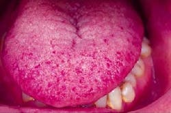
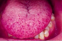
The patient's vital signs were all found to be within normal limits. Examination of the head and neck region revealed no enlarged or palpable lymph nodes. Examination of the soft tissues of the oral cavity revealed multiple, tiny, and red pin-head-sized spots on the tongue, buccal mucosa, and lips (see photograph). Diascopy revealed that the red spots easily blanched with pressure.
Clinical diagnosis
Based on the clinical information available, which one of the following is the most likely diagnosis?
- Peutz-Jeghers syndrome
- Darier's disease
- Hereditary hemorrhagic telangiectasia
- Focal epithelial hyperplasia
- Idiopathic thrombocytopenia purpura
Diagnosis
- Hereditary hemorrhagic telangiectasia
Discussion
Hereditary hemorrhagic telangiectasia (HHT), which is also known as Osler-Weber-Rendu Syndrome, is a genetic disease that is inherited as an autosomal dominant trait. It is an uncommon disorder that affects both the skin and mucous membranes.
Clinical features
HHT is characterized by the abnormal vascular dilation of the terminal vessels. The term telangiectasia refers to the dilation of widening (-ectasia) of a blood vessel (-angi) at its furthest or terminal point (tel-).
The telangiectatic vessels appear clinically as red macules or papules on the skin as well as the oral and nasal mucous membranes. Such lesions appear early in life. After puberty, the size and number of the lesions tend to increase with age.
The telangiectatic vessels blanch with pressure. This indicates that the red color is due to the blood contained within the capillaries that are close to the surface of the mucosa. Skin areas of involvement often include the face, chest, hands, and feet. Although any intraoral area may be involved, the most common locations include the vermilion of the lip, tongue, and buccal mucosa.
Lesions that involve the nasal mucosa may cause epistaxis or nose bleeds. Epistaxis is the most common presenting sign of HHT. If the mucosa of the gastrointestinal tract is affected, bleeding may occur. Control of such mucosal bleeding may be problematic and, if severe, may result in anemia. Other complications include the formation of arteriovenous fistulas in the lungs, liver, and brain. The brain lesions may predispose the patient to the formation of brain abscesses.
Diagnosis and treatment
The diagnosis of HHT is made based on the clinical findings, the history of mucosal bleeding, and family history.
Mild cases of HHT may require no treatment. In cases where bleeding is a moderate problem, cryosurgery or electrocautery of the involved telangiectasias may be indicated. In severe cases of epistaxis, a surgical procedure of the nasal septum may be required.
Some authors recommend the use of antibiotic prophylaxis in patients with HHT prior to any dental procedures that may induce bacteremia. Such antibiotics are recommended because of the risk of brain abscess formation.
The prognosis of a patient with HHT is good, with a 1 to 2 percent mortality rate associated with blood loss. For patients with HHT who develop brain abscesses, the mortality rate is 10 percent.
Joen Iannucci Haring, DDS, MS, is an associate professor of clinical dentistry, Section of Primary Care, The Ohio State University College of Dentistry.

