Gingival cyst of the adult

by Nancy W. Burkhart, BSDH, EdD
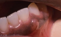
The patient is 49-year-old Lewis who has entered your practice today for a routine visit. He is being treated for hypertension, consumes alcohol in moderation, and is a nonsmoker as of his last appointment. When you ask him if he has noticed any changes in his body, including his mouth, he points to the enlarged area in the lower left quadrant (see Figure 1). He has noticed this “bump” and is especially aware of it when he flosses.
He asks you what it could be, and is it cancer? He is very apprehensive and indicates that this is really the reason he has come in to see you. You find out that he has recently been to a program on lifestyle changes at his company, and they talked about smoking and oral cancer.
The patient was given an oral cancer screening, radiographs, and was referred to an oral surgeon/oral medicine specialist for a biopsy of the area in question.
The diagnosis after a biopsy was the gingival cyst of the adult.
Etiology: The gingival cyst of the adult (GCA) is considered a variation or counterpart to the lateral periodontal cyst. GCAs are developmental and noninflammatory, as well as rather uncommon with reports ranging from .5 to 1% of reported odontogenic cysts.
The cyst usually occurs in the fourth through sixth decade of life, although a wide age range from 14 to 84 has been reported in research findings. The mandible is affected 60% of the time, and some have reported a slight female predilection (Kelsey, 2009). The lesion is usually less than 1.0 cm in diameter (Wysocki et al. 1980). The favored location is the mandibular canine-premolar region.
Pathogenesis: The cyst originates from the dental lamina and the stimulation of epithelial rests (rests of Serres) that is thought to cause the development of the cyst. Explanations include that the GCA originates from the epithelial connection between the mucosa and the enamel organ, and the remnants of the cell rests of Malassez. It is believed that this cyst of the adult in the soft tissue is the counterpart of the lateral periodontal cyst that exists in bone.
Perioral and intraoral characteristics: The gingival cyst appears as a nodule that is painless, smooth, nonblanching, and may be normal in color. The area is usually nonmobile, flesh-colored with a light blue hue in some cases, and a firm nodule that may be slightly depressed with pressure.
Distinguishing characteristics: The tooth will test vital unless prior damage has occurred to the pulp from other sources. The GCA will usually not appear radiographically distinct unless the cyst is large and emits pressure on the bone, creating resorption leading to a depression (see Figure 2).
Significant microscopic characteristics: The lining of the cyst may be squamous, cuboidal, or columnar with usually three layers of cells. The specimen will have clear cells of glycogen-rich cytoplasm within the tissue that are deemed a diagnostic feature of GCA. It is important to provide a full histological examination of the lesion.
Dental implications: The gingival cyst of the adult may be misdiagnosed as a mucocele because of the similar clinical appearance in some cases. Mucoceles often have a light, milky blue color, and this is sometimes also found with the gingival cyst of the adult. The mucocele is often more flacid, moveable, and not firm compared to the GCA. In addition, the mucocele occurs in areas that have salivary tissue that is stimulated.
Therefore, a mucocele on the gingiva would be rare. A clinical differential diagnosis would be:
- Fibroma
- Peripheral odontogenic fibroma,
- Pyogenic granuloma
- Peripheral giant cell granuloma
The latter two should be considered when there is not a vascular component.
Treatment and prognosis: Treatment is surgical excision and histopathologic examination for a conclusive diagnosis. Defects in the bone may be observed when the growth is removed. Again, the defects are often not detectable on a radiograph.
As always, keep listening to your patients and asking good questions.
References
Bell RC, Chauvin PJ, Tyler MT. Gingival cyst of the adult: a review and report of eight cases. Can Dent J. 1997; 63(7):533-535.
Kelsey WP. Kalmar JR, Tatakis DN. Gingival cyst of the adult. Regenerative therapy of associated root exposure. A case report. J Periodontol 2009; 81(12): 2073-81.
Jones AV, Craig GT, Franklin CD. Range and demographics of odontogenic cysts diagnosed in a UK population over a 30-year period. J Oral Pathol Med 2006;35(8); 500-507.
Ojha J, Gupta A, Kwapis-Jaeger J. Oral pathology quiz #21. Gingival cyst of the adult. J Mich Dent Assoc. 2010;92(5): 28, 30.
Wysocki GP, Brannon RB, Gardner DC, et al. Histogenesis of the Lateral Periodontal Cyst and the Gingival Cyst and the Gingival Cyst of the Adult. Oral Surg Oral Med Oral Pathol. 1980; 50:327-334.
Past RDH Issues