Pain Control: The Options
by Laura J. Webb, CDA, RDH, MS
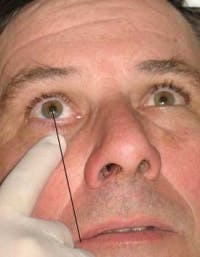
Practicing hygienists often share with me that they avoid providing certain maxillary injections due to a variety of concerns, including clinician confidence and patient safety and comfort. Many concerns may be alleviated by careful review of the step-by-step techniques provided in textbooks and literature and considering the tips provided below.Fig. 1: 45 degrees to occlusal and midsagittal (see line) planes (All photos courtesy of LJW Education Services)
For all maxillary injections (except palatal), a prerequisite to success includes drying the tissue and making/keeping it taut. Doing this facilitates effective, painless penetration and good vision during advancement of the needle. When the tissue is allowed to collapse around the needle, it impairs our ability to see the needle movement and we may not advance the needle adequately. In some situations, the use of a 2x2 cotton gauze may help make retraction easier.
Posterior superior alveolar nerve block (PSA)
I find the PSA one of the essential injections to be able to provide my patients, particularly when performing quadrant/sextant debridement procedures. I like it because it has a high success rate for profound and sustained anesthesia to the molar pulps and buccal soft tissues with one injection and because it is comfortable for the patient. I find this to be consistent whether I choose lidocaine, prilocaine, or articaine. The alternative, local infiltration in this area, often requires multipleAnterior superior alveolar nerve block (ASA)
When I was in school, we gave a local infiltration above the cuspid and called it the ASA. Not so! For the “true” ASA (sometimes referred to as the infraorbital nerve block), insertion is usually over the first premolar and deposition near the area of the infraorbital foramen. Although the literature supports that following properPalatal injections
Dental hygienists often need palatal anesthesia during the provision of care, and yet many are reluctant to provide it because they fear it will be uncomfortable for the patient. The provision of palatal injections should be perfected, not feared. They can be atraumatic if proper techniques are utilized. The key components for administration of successful and atraumatic palatal anesthesia with a traditional syringe and needle include: 2,3,4,6- Clinician confidence
- Adequate topical (two minutes)
- Pressure anesthesia
- Prepuncture technique/“anesthetic pathway” (Fig. 3a)
- Maintain control of needle (27 gauge short)
- SLOW deposition
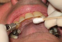
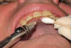
Knowing that I can provide an atraumatic palatal injection is important to actually producing that desired outcome. And although the use of topical anesthetic may be controversial for palatal injections due to the keratinized nature of the hard palatal tissue, I find that taking the time to let the topical anesthetic do its job is worth the wait. The use of pressure anesthesia is very important for patient comfort. The cotton swab (avoid the use of a mirror handle as it can be very uncomfortable for the patient) placed strategically with very firm pressure next to the insertion site works well. The prepuncture technique (Fig. 3a) involves bowing the needle and placing a few drops of anesthetic on the tissue, prior to penetration.3,4,6 Advancing the anesthetic solution ahead of the needle provides an “anesthetic pathway”4, which aids in a more comfortable deposition.
Nasopalatine nerve block (NP)
The NP nerve block is perhaps the most avoided of all palatal injections. Traditionally the NP injection has been provided using the techniques listed above with the insertion site next to the incisive papilla (Figs. 3a,b). The most common error in providing this injection is the insertion of the needle directly into the incisive papilla, which is very painful for the patient. In addition, clinicians sometimes attempt to inject too rapidly and at a shallow depth.
An option to the traditional approach to the NP nerve block that many hygienists employ is the technique whereby multiple penetrations prior to the palatal penetration are performed. This technique takes longer but is time well spent. The first injection is into the labial frenum, anesthetizing the facial soft tissues in the area, the second into the already anesthetized interdental papilla between the central incisors, and the third into the palate next to the incisive papilla. Because the palatal tissue should be anesthetized by the second injection, topical anesthetic, pressure anesthesia, and anesthetic pathway techniques usually are not required for the third injection (palatal) (Figs. 4a,b,c).
Fig. 5a: Locating anterior foramen Fig. 5b: Insertion site
Anterior palatine (greater palatine) nerve block (AP)
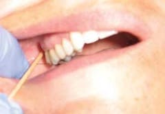
One challenge that clinicians report facing with the provision of the AP nerve block is locating the anterior foramen. The anterior foramen may be located by using a cotton swab to firmly palpate from the first molarFig. 5c: Fulcrum (no swab shown)
Local anesthesia, when needed, is an important service we provide for our patients. Learning strategies to increase our confidence, knowledge, and improve our technique is a professional, lifelong process. We should continuously evaluate our strengths, biases, and challenges and incorporate evidence-based strategies for delivery of local anesthesia to facilitate our professional growth and improve the quality of care we provide.
Laura J. Webb, CDA, RDH, MS, is an experienced clinician and educator who owns LJW Education Services, www.ljweduserv.com. She provides educational methodology courses and accreditation consulting services for DH/DA education programs and CE courses for professionals. Laura frequently speaks on the topics of local anesthesia and nonsurgical periodontal instrumentation.
References
- Blanton P, Jeske A. Avoiding complications in local anesthesia induction — anatomical considerations. J Am Dent Assoc, 2003; 134(7):888-893.
- Darby M, Walsh M. Dental Hygiene Theory and Practice 2nd ed., 2003; Saunders.
- http://www.fice.com/course/FDE0010/c12/p01.htm Accessed 1/4/10.
- http://www.milesci.com/compudent_id.html Accessed 1/4/10.
- Malamed S. Complications in local anesthesia administration. Dimensions of Dental Hygiene, Oct. 2006; 4(10):28-33.
- Malamed S. Handbook of Local Anesthesia 5th ed., 2004; Elsevier.
- Freuen N, Feil B, Norton N. The clinical anatomy of complications observed in a posterior superior alveolar nerve block. The FASEB Journal 2007; 21:776.4.
