Panning into the future: A wide angle view of intraoral cameras helps determine your needs
The evolution of intraoral cameras means a wide angle view when determining which one best suits your needs
By Donna Lesser, RDH, EdD
Over the last 10 years, digital imaging has taken on an important role in the process of patient care in dentistry. Companies are now producing digital intraoral cameras that are easier to use and multifunctional. These offer higher image quality and imaging software that integrates seamlessly with patient management systems.
While these advances have made the intraoral camera one of the most valuable pieces of dental equipment, they have not made it any easier for clinicians to find an answer to the decades-old question, "Which camera is right for my practice?" Instead, they have created new questions for clinicians to consider: Should the practice update its intraoral cameras to maximize the usage of digital images during patient care? And how many cameras does the practice require to meet its needs?
The right intraoral camera
Without question, determining what the camera will be used for and what images will be captured is the most important step in selecting the right intraoral camera for the practice. This includes considerations of what the camera will be used for today, as well as what it will be used for over the next three to five years.
Will it be used to capture images for documentation purposes only? Will the images be used for case presentations to patients or for referrals to specialty practices? Will they be used for documenting shade to be shared with the lab or to record the results of in-office whitening procedures? Perhaps the only intended use will be for oral hygiene and patient education. In the future, is there a possibility the intraoral camera will be used for teledentistry?1 Each of these uses may require a slightly different image quality.
Another important consideration is determining the dimensions of the area the intraoral camera image will capture. Will it capture images of a single tooth, tooth surface, or lesion? Or will it be used for larger areas, such as a quadrant or even extraoral images? And lastly, will there be a need for video capturing? Having a vision for how the camera and images will be used will assist with finding the right intraoral camera.
Image quality
The quality of the image should match the camera's intended or projected use. Most intraoral cameras have built-in autofocus abilities that make capturing an image easier for all users. An autofocus function can enhance the quality of the images being captured. If a camera does not have autofocus capabilities, it will have a sliding bar that can be used to focus the image. Both enable the clinician to maximize the quality of the image. Cameras such as the USBCam4 from Sirona Dental offer a fixed focal range and one-click image capture, which can help when multiple images of the same area need to be taken quickly to minimize patient time at the office.
In addition to focusing capabilities, consideration should also be given to the optics, resolution, lighting, depth of field, and glass lens elements of the camera.
Optics-An intraoral camera's optics has a profound impact on image quality. Intraoral cameras have either a CCD (charge-coupled device) sensor or a CMOS (complementary metal-oxide semiconductor) sensor, which is located in the handpiece.2 Both receive light and convert it into an electronic signal that is processed through the camera's imaging software to produce an image on the computer monitor.
The location of the CCD chip or CMOS sensor has an impact on the quality of image that is produced; a higher-quality image is produced when the sensor is placed at the end of the handpiece near the lens. If it is placed farther from the lens, it will produce an additional prism that degrades the quality of the image. This degradation of the image might or might not be relevant, depending on what the images will be used for.
Resolution-Resolution, or the amount of detail that the intraoral camera can capture in an image, is measured in pixels. In theory, a camera with more pixels will take more detailed images that can be displayed at larger sizes on the computer screen without being blurred.2 With intraoral pictures, more detail allows for a more accurate recording of the object being photographed. Therefore, higher-resolution cameras are highly recommended if image quality is a top priority.
Manufacturers will provide the intraoral camera's resolution for both still images and video if the camera performs both functions. Camera resolutions currently range from 640 pixels wide by 480 pixels high to 1,280 pixels wide by 720 pixels high. To determine what fits the needs of your practice, get a demonstration of more than one intraoral camera to evaluate the quality of the images with different resolutions.
Lens-In an attempt to differentiate their intraoral cameras from others on the market, more manufacturers are disclosing the elements of their cameras' lenses. An "element" is individual glass lens within the lens. More elements can add to a camera's capability to control optical defects or enhance the image. Unfortunately, at the same time, more elements can cause a lower image contrast. Currently, the consensus is that the number of elements doesn't make a lens superior or inferior with respect to the quality of the image.
Instead of a spherical lens, an aspherical lens is being used in some intraoral cameras to increase the sharpness of the images being captured. Unlike a spherical lens, an aspherical lens can optically correct the focus of the LED lights to a single point. Aspherical lenses have been used for years in traditional camera lenses, and many claim that this feature is regarded as the mark of quality. We have yet to determine if these lenses will have the same effect on intraoral images.
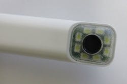
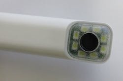
LED lighting-LED lighting provides a continuous source of light to eliminate the need for a flash. LED lighting has also replaced the halogen bulbs that were first used with intraoral cameras. More LEDs yield a higher light output, which provides better images. Intraoral cameras typically have four to eight LEDs; those with six or more LEDs produce more accurate color contrast and images. An example of a system with high clarity and color due to its eight LEDs is Sirona's USBCam4 (see Figures 1, 2).
Figure 1
Figure 2
Depth of field-The depth of field is basically the range of distance from the camera to the object that allows the image to appear sharp. The image's sharpness changes gradually as the camera changes distance from the object, but the variation in sharpness is noticeable when a camera is moved outside of its depth of field.3 Cameras that have a long depth of field can be moved closer to or farther from the object while keeping the image on the computer screen in focus.
An example of a long depth of field for an intraoral camera would be a range up to 45 mm. With a short depth of field, the image will become unfocused during the changing of the camera distance in relationship to the object. If the only use of the intraoral camera is for patient education or video capturing, the need to have the image remain focused is essential. Some cameras have up to three focal ranges and a depth of field up to 130 mm, which allows them to take both intraoral and extraoral images.
Barriers-All intraoral cameras need to have barrier sheaths on them during usage to prevent cross-contamination. If the barrier sheath does not fit the handpiece snugly, the image that is being captured will be distorted due to wrinkles or lines from the barrier. To eliminate the image distortion that can occur when a poor-fitting barrier sheath is used, many manufacturers develop barrier sheaths specifically for their intraoral cameras.
If your practice has several intraoral cameras, look for a company that manufactures barriers designed to adjust to fit multiple intraoral cameras. It is also recommended to place a barrier sheath on the intraoral camera to see its effect on the image before purchasing the camera.
Design and ease of use
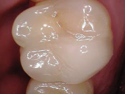
Intraoral camera designs have become lighter and more balanced over the last 10 years, which has helped to reduce operator fatigue (see Figure 3). The weight of the handpieces can vary, but all are ultra-light and each can be compared to the weight of two to three slices of bread. Some manufacturers claim their cameras are balanced so that the operator will feel the weight distributed equally in his or her hand. It is recommended to evaluate several cameras to determine if there is a difference in the weight distribution and, if so, if it makes a difference.
Figure 3
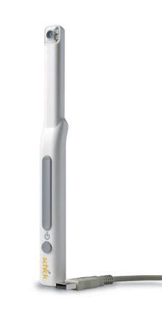
Figure 4
Whether barrier sheaths will fit is also an important element of a camera's design. As previously noted, custom or adjustable barriers, as opposed to generic barriers, are recommended to eliminate image distortion. The intraoral camera must also be able to withstand being cleaned with an EPA-registered (intermediate-level) disinfectant after each patient for whom it's used.
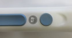
Most intraoral cameras are activated automatically when lifted out of the holder and are deactivated when returned to the holder. A few cameras have a power button located on the handpiece to control the camera (see Figure 4). Additionally, most intraoral cameras have a one-button/one-click image-capturing mechanism. Those with the capability for live-video display have a one-button freeze-and-capture mechanism. These mechanisms allow the operator to focus on the image being captured and not on how to capture the image.
Intraoral cameras are now considered portable, as most have a "plug-and-go" feature. They can easily be connected to the computer via a USB adapter or wirelessly.4 This feature allows the cameras to be carried and used between multiple operatories. The USB-connected cameras, such as Sirona's camera, do not require a capture card since the USB cord connects them directly to the computer. When considering purchasing this type of camera, consider whether the cord is long enough to reach from the computer to the patient. The cordless intraoral camera designs do not have distance limitations and are equally convenient to move between operatories.
In general, an intraoral camera should not need routine maintenance if it is handled and cared for according to the manufacturer's guidelines. The life span of an LED light is approximately 500,000 hours, which translates into multiple years. Intraoral cameras' dependability is an amazing feature that is often taken for granted by users.
Other considerations
In addition to the aforementioned features and capabilities, there are other considerations to be aware of during the selection process. These include compatibility of the imaging software with the practice's patient management system, computer system requirements to support the software, and support services provided by the manufacturer.
Patient management system-Prior to purchasing an intraoral camera, make sure the camera's software is compatible with the practice's patient management system. Most cameras' software programs are compatible with Dentrix, DEXIS, and Eaglesoft; however, all manufacturers will provide the software's capability specs. Find out how the images are imported into the patient chart. Are the images seamlessly imported, or are steps required to get a digital image into a chart? An intraoral camera with imaging software that integrates easily into your patient management system is highly recommended.
Computer system requirements-The intraoral camera's imaging software must be compatible with the computer(s) that the camera will be used on. All are compatible with Microsoft Windows, and some are also compatible with Mac OS. Imaging software can require up to 1 GB of RAM space and can work with a 32-bit processor, a 64-bit processor, or both; therefore, computer systems may need to be upgraded to support the imaging software. Purchasing a camera with imaging software that is compatible with both 32- and 64-bit processors will provide more flexibility with future computer upgrades.
Support services-In general, intraoral cameras come with a 12- to 34-month warranty. Although intraoral cameras are easy to use, purchasing one from a manufacturer that offers user-support services is highly recommended. Integrating the imaging software with your patient management system will go more smoothly with tech support from the manufacturer. The computer skills and comfort level of the users will determine the extent of the support services needed for the practice.
Cost-Intraoral cameras can range from the low hundreds into the thousands of dollars. Simply put, having an intraoral camera that provides quality images and meets the needs of the practice outweighs the cost of the equipment. There is no question about it: You get what you pay for.
There is no one-size-fits-all approach to finding an intraoral camera since every practice has different needs. Prioritize the needs of your practice, determine what features are negotiable, and begin by evaluating cameras at dental meetings or by requesting in-office demonstrations. During the selection process, remember that intraoral cameras are underutilized-think about how you'll use yours to enhance patient care both now and in the future. RDH
Donna E. Lesser, RDH, EdD, is a full-time associate professor in the dental hygiene program at Moreno Valley College in California. She began working at Moreno Valley College in 2002, following 21 years of clinical practice and seven years in both dental and dental hygiene education. Dr. Lesser has a doctorate in teaching technologies from Pepperdine University. Her interests include integrating technology into education and clinical practice, educational methodology, periodontology, and research. Dr. Lesser can be reached at [email protected].
References
1. Inês Meurer M, Caffery LJ, Bradford NK, Smith AC. Accuracy of dental images for the diagnosis of dental caries and enamel defects in children and adolescents: A systematic review. J Telemed Telecare. 2015;21(8):449-458.
2. Desai V, Bumb D. Digital dental photography: A contemporary revolution. Int J Clin Pediatr Dent. 2013;6(3):193-196.
3. Terry DA, Snow SR, McLaren EA. Contemporary dental photography: Selection and application. Compend Contin Educ Dent. 2008;29(8):432-436.
4. Feuerstein P. Take a look at me now. Dent Today. 2015;34(5):34.
