Oral lesion documentation: Dental patients could start monitoring own progress during treatment
Patients could start to monitor their own progress during treatment
By Nancy W. Burkhart, BSDH, EdD
Dentists and hygienists are continually documenting the progress of their patients, recording lesion findings or changes in the tissues of these patients. For many years, a handwritten paper-chart entry was considered the only form of legal documentation for oral findings. With new technology and improvements in computer technology, a majority of facilities have gone paperless with computerized documentation.
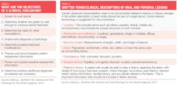
Individuals have become quite sophisticated in documenting their own health issues, even adding images that they take with their cell phones. According to a John Hopkins study (2016), medical mistakes are the third most common cause of death, and patients are realizing the importance of their own active role in overall health (see Tables 1 and 2).
An opinion essay written by Dr. Leana S. Wen (July 2016), who is an emergency physician and commissioner of health in Baltimore, presents a perspective about the way health care might have evolved by the year 2066. She states that "we need to accelerate the movements to have patients and doctors as shared partners in medical decision-making." She adds that by 2066, "In real time, you and your care team agree on your treatment plan."
She goes on to say that, in 2066, a lack of access to your own medical records is a thing of the past. The essay states that you will be responsible for much of the documentation along with your doctors and your advocate that has been assigned to you. Doctors will come to you, when it's good for your schedule. Technology will be very advanced, and some diagnostic features will be performed from a distance with information relayed to the physician through handheld devices. Innovation is certainly in the air. Within the span of the next 50 years, who knows?
We have been conducting the International Oral Lichen Planus Support Group since 1997. At one time, few patients sent us e-mail material with images, but we have seen a significant increase in this combination during the past couple of years. We hear from patients worldwide who are searching for information regarding oral conditions and specifically, oral lichen planus. Often, patients will provide a complete list of previous visits to health-care providers, and sometimes they will send digital images of what they are concerned about in their mouth or skin. Sometimes, patients may even send a series of progressive images of the lesions that they have taken themselves over time.
Figure 1: This image was taken with an iPad Mini. Courtesy of Dr. Thomas Toffoli in San Ramon, CA.
Patients have become very proficient with the latest technology. The age range of patients with lichen planus is usually an older age group, who seem to do quite well with new technology.
Many clinicians work in offices that have intraoral camera devices, and these devices take excellent images of oral lesions. Descriptions, measurements, and oral lesion documentation of findings will always be legally needed. But the old saying that "a picture is worth a thousand words" still rings true.
I spent two years in a postdoctoral study in the oral pathology unit of a university many years ago. The two oral pathologists were always delighted when the practitioner included an image of the lesion along with the tissue specimen. This image just added another dimension to their diagnostic tools as they viewed tissue under the microscope. The image itself was not diagnostic, but it certainly provided an insight into the actual patient profile. Accurate written descriptions are very important for any type of lesion, whether in the mouth or skin.
Patient Documentation
Most patients have a cell phone with them during their appointment, so the clinician can take an image of the lesion (at least those that are not in the very posterior area of the mouth) with the patient's cell phone. Since these returning patients are seen at least once a year, clinicians can pull up the image, and it can be viewed during the next appointment and compared to its present state. The patient then has access to compare changes that may occur and to check any progress when using oral medications for treatment of the lesions. The quality of the cell phone image has become quite good and readily available. We liked this idea presented and think that it adds even more involvement with the patient in their own health-care management. Of course, for some lesions or tissue changes that need a very close, diagnostic view, intraoral camera use is always best when available.
Cell phones and tablets take great images, and the patient can send these via e-mail or retain for future comparisons and documentation (see Figure 1). The camera quality with flash provides a sharp, detailed photo. Many devices are available to attach to tablets and cell phones that promote very close views of tissue. Some phone cameras will be of higher quality than others.
Some clinicians say that it takes time to set up a camera or to connect those associated with their computers. Images taken using an in-office device such as intraoral cameras in the dental office or clinic facility provide another way of documenting changes and should be part of the patient's permanent record. Cell phone/tablet photography is certainly quick and easy to do when the area in question is accessible to reach with the device. Again, this is another tool. I think that patients are constantly trying to assess whether oral medications are improving their tissue, and this does assist in a comparison of tissue changes.
The ratings of cell phones are based on multiple factors, including the megapixels, flash, optical stabilization, front and rear cameras, panoramic modality, video quality, motion, and time lapse. The 12- and 13-megapixel quality seems to be the optimal range. Each year, new improvements occur with the technology, so it is wise to evaluate the importance of your phone and if the camera is an important feature to you.
An Oral Medicine Perspective
The continued monitoring of patients who have chronic long-term disease states is extremely important. Skin diseases such as lichen planus, pemphigus, and pemphigoid are chronic, long-term diseases. With the use of cameras, computers, and cell phones, patients learn to document their intra- and extraoral reaction (both positive and negative) to treatments such as medications, possible trigger mechanisms, nonhealing tissue, and new areas of concern.
Sollecito (2013) stated: "Oral medicine is often regarded primarily as a nonsurgical dental specialty that includes diagnosis, physical evaluation, and therapeutic management of medically related oral disorders. These disorders include salivary gland disease, oral complications resulting from systemic disease, chemosensory and neurologic impairment of the oral and maxillofacial complex, orofacial pain disorders, temporomandibular disorders, and oral mucosal diseases."
This goal of patient management of medically related oral disorders continues to be an integral part of total patient care. Monitoring and documentation play a huge role in successful patient management, and patients have better outcomes when they are involved in their own health care treatments.
As always, continue to ask good questions and always listen to your patients. RDH
References
1. Consumer Reports: http://consumerreports.org
2. DeLong L, Burkhart NW. General and Oral Pathology for the Dental Hygienist. Wolters Kluwer/Lippincott Williams & Wilkins. Baltimore, 2013.
3. Sollecito TP, Rogers H, Prescott-Clemons L, et al. Oral medicine: Defining an emerging specialty in the United States. Journal of Dental Education, 2013; 77:4, 392-394.
4. TechAdvisor: pcadvisor.co.uk/feature/mobile-phone/best-phone-camera-of-2015-test-3630896/
5. Wen LS. A new vision of true health. Consumer Reports. July 2016. 81:7;52-53.
6. McMains V. http://hub.jhu.edu/2016/05/03/medical-errors-third-leading-cause-of-death/
NANCY W. BURKHART, BSDH, EdD, is an adjunct associate professor in the department of periodontics/stomatology, Baylor College of Dentistry and the Texas A & M Health Science Center, Dallas. Dr. Burkhart is founder and cohost of the International Oral Lichen Planus Support Group (http://bcdwp.web.tamhsc.edu/iolpdallas/) and coauthor of General and Oral Pathology for the Dental Hygienist. She was awarded a 2016 American Academy of Oral Medicine Affiliate Fellowship (AAOMAF). She was a 2006 Crest/ADHA award winner. She is a 2012 Mentor of Distinction through Philips Oral Healthcare and PennWell Corp. She can be contacted at [email protected].


