Probe selection: A hygiene team's review of periodontal probes considers several factors

By Yemia A. White, RDH, Cheri K. Robinette, RDH, and Amy Bahry, RDH
Does this scenario sound familiar? You just completed a continuing-care appointment, which included a comprehensive periodontal evaluation, and the doctor enters your room to examine the patient.
The doctor proceeds to spot probe a few areas and asks, “What did you probe on the mesiobuccal of tooth number 15?”
You review your periodontal chart and confidently reply, “5mm’s with bleeding on probing.”
After a brief pause, the doctor replies, “Actually, I am probing, 6.5 almost 7mm’s.” Bemused, you lean in for a better visual, and can now clearly see what the doctor sees. You acknowledge the discrepancy, but wonder why your measurements are so different from the doctors.
Background
As a chronic infection, periodontal disease affects 47.2% of the adult U.S. population aged 30 and older.(1) Gingivitis is caused by bacterial biofilm accumulating on teeth adjacent to the gingiva.(2) Gingiva that is affected by gingivitis typically appears red, swollen, and shiny. It may be tender and bleed easily during brushing and flossing. Fortunately, this early stage of infection only affects the gingiva and can be resolved with regular dental visits and proper oral hygiene techniques.
On the other hand, the more aggressive periodontitis results in the loss of connective tissue and bone support.(2) Periodontitis is categorized in three stages: slight (mild), moderate, and severe (advanced).(3) Moderate and advanced periodontitis may be easier to diagnose due to apparent clinical and radiographic evidence such as bone loss, swollen and bleeding gingiva, loss of papillae, deeper pocket depths, gingival recession, clinical attachment loss, tooth mobility, and malodorous breathe.
In comparison, slight periodontitis is more ambiguous—often causing subtle, isolated changes in the periodontium that are less clearly defined, which may result in the misdiagnosis or mistreatment of the disease.
In addition to oral health concerns, research regarding the connection between oral health and systemic health has confirmed that diabetes increases the risk of developing periodontal disease.(4) Increasing evidence supports the role of systemic inflammation and its relationship to periodontitis and cognitive decline.(5) Ongoing research continues to explore the association between inflammation and diseases such as hypertension,(6) cardiovascular disease,(6) and rheumatoid arthritis.(7) Accordingly, the oral-systemic connection substantiates the need for thorough periodontal assessments that encompass accurate data collection through careful observation and skill.
The Importance of Probing
Periodontal probing is one of the most common methods used in diagnosing periodontal disease.(8) Regular use of periodontal probes in routine dental practice facilitates and increases the accuracy of the process of diagnosing the condition, formulating the treatment, and predicting the outcome of therapy.(9) Although critical for accurate diagnosis, baseline assessments, and legal documentation, periodontal probing may be met with resistance from patients and clinicians. Remarks from patients such as “I hate this part” or “Of course my gums are going to bleed when you stick them” are red flags that the patient may associate probing with pain or “being poked.” This, in turn, can result in patients being apprehensive about probing, refusing periodontal exams, and being unreceptive to treatment recommendations—ultimately leaving infection untreated.
In contrast, dental hygienists may be resistant to comprehensive periodontal evaluations where probing depth, bleeding indices, gingival recession, attachment loss, furcation involvement, and mobility is recorded. Common pitfalls for dental hygienists are habitual “spot probing” or eliminating probing altogether due to a shortage of time, lack of assistance, patient discomfort, and poor visibility.
While periodontal probing is the primary means for measuring pocket depth and clinical attachment, many variables can affect its outcome. Various factors influence the extent of probe penetration: gingival health, probing force and examiner variability, probe design, and obstruction factors such as crown contours and calculus.(10)
Even though variance with probing results has been well documented, it is still important to try to minimize discrepancies whenever possible. Several methods help minimize inconsistencies in probing. First, using a hands-free or voice-activated system will help reduce errors in determining the state of active periodontal disease.(9) Secondly, wearing magnification lenses will improve posture and enhance indirect vision skills,(11) which helps increase efficiency and accuracy. Third, using the same type of probe (especially if there are multiple hygienists in the practice) and selecting one that is easy to read will improve consistency among clinicians. Finally, selecting a probe that reduces patient discomfort will help increase patient compliance and relieve apprehension associated with probing.
Types of Probes
Periodontal probes are classified into three categories. First generation or conventional probes are calibrated and consist of a handle, a shank, and tip.(12) The tip is rounded with different types of gradations marked in millimeters.(12) Second generation probes are pressure-sensitive,(12) and third generation probes are computerized probes that are extremely accurate but also expensive.(12)
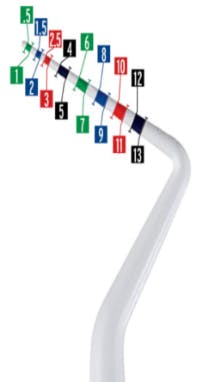
Are dental hygienists as meticulous about probe selection as they are about scalers and curettes? If not, why? An optimal probe should meet specific criteria. The new Colorvue probe (Goldstein Colorvue Probe by Hu-Friedy) has been created with precision, accuracy, visibility, and patient comfort in mind. The probe is designed in mm increments of .5, 1, 1.5, 2, 2.5, 3, 4, 5, 6, 7, 8, 9, 10, 11, 12, and 13 for increased accuracy, especially in measuring sulcular depth for margin placement (see photo above). The probes’ tip is fabricated with a medical grade resin that is demarcated in vivid colors for improved visibility.
Survey
In order to evaluate whether or not the new Colorvue Probe should have a place in our periodontal evaluations, 100 patients were tested to compare the Colorvue probes with the black color-coded metal probes we were using. Our goal was to determine if patients noticed a difference between the two probes. In addition, each hygienist completed an evaluation of the use of the new probe.
The survey’s questions focused on the probe’s ease of use, accessibility to areas being checked in the mouth, and accuracy. Before probing, all patients were asked to evaluate each probe based on how it felt to the gingiva. Each hygienist probed three teeth with one of two probes. The same teeth were re-probed with the other instrument, and patients were asked if they noticed a difference between the two.
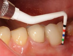
Overall, the hygienists agreed that the Colorvue probes’ lightweight and ergonomically correct handle-size were positive attributes. All reported that the vibrant color-coded demarcations improved visibility when compared to a metal probe with black color-coding (see photo above).
Similarly, the hygienists found the narrow, flexible tip allowed for easier access around natural teeth and implants. Finally, 61 of the 100 patients surveyed favored the Colorvue Probe over the metal probe, citing it was gentler to the tissue.
Currently, probing is the standard used to measure pocket depth. It is a critical component in the overall assessment of periodontal health. Although imperfect, manual probing is still widely used for periodontal probing. Based on the positive feedback we received from our patients, we have implemented the Goldstein Colorvue Probe into our periodontal assessments. We are optimistic that, with its continued use, more patients will be less apprehensive about probing and more accepting of routine periodontal probing.
Yemia A. White, RDH, is a practicing dental hygienist for Goldstein, Garber, and Salama, LLC. She is a member of ADHA and can be reached at [email protected]. Cheri K. Robinette, RDH, joined Goldstein, Garber, and Salama in 2003. She is a member of the Association of Dental Implant Auxiliaries. Amy L. Bahry, RDH, also joined Goldstein, Garber and Salama in 2003 and is also a member of the Association of Dental Implant Auxiliaries.
References
- Smiley C, Tracy S, Abt E, Michalowicz B, John M, Gunsolley J, et al. Systematic review and meta-analysis on the nonsurgical treatment of chronic periodontitis by scaling and root planning with or without adjuncts. ADA Center for Evidence-Based Dentistry. 2015;1-179.
- Philstrom B, Michalowicz B, Johnson N. Periodontal diseases. The Lancet Seminars. 2005;366(9499):1809-1820.
- American Academy of Periodontology (AAP). American Academy of Periodontology task force report on the update to the 1999 classification of periodontal diseases and conditions*. 2015;86(7):835-838.
- Mealy B. Periodontal Disease and Diabetes: a two-way street. JADA. 2006;137:26s-31s.
- Ide M, Harris M, Stevens A, Sussams R, Hopkins V, Culliford D, et al. Periodontitis and Cognitive decline in Alzheimer’s disease. (2016). PLoS ONE 11(3):e0151081. doi:10.137/journal.pone0151081.
- Angeli F, Verdecchia P, Pelligrino C, Pelligrino R, Proscuitti L, et al. Association between periodontal disease and left ventricle mass in essential hypertension. Hypertension. 2003;41:488-492.
- Kaur S, White S, Bartold PM. Periodontal disease and rheumatoid arthritis. J Dent Res. 2013;92(5):399-408.
- Garnick JJ, Silverstein L. Periodontal probing: probe tip diameter. J Periodontal. 2000;71 (1):96-103
- Ramachandra SS, Mehta DS, Sandesh N, Baliga V, Amarnath J. Periodontal probing systems: a review of available equipment. Compend Contin Educ Dent. 2011;32(2):71-7.
- Atassi F. Periimplant probing: positives and negatives. Implant Dentistry. 2002;11(4):356-362.
- Hoerler B, Branson B, High A, Mitchell T. Effects of dental magnification lenses on indirect vision: a pilot study. The Journal of Dental Hygiene. 2012;86(4):323-330.
- Martu A, Popa C, Luchian L. A., Martu I, Oanta C, Martu S. Evaluation of the efficiency of 2 types of periodontal probing. Balk J Dent Med. 2015;19:163-166.