A 25-year-old female visited a dentist for an initial examination. Radiographic examination
A 25-year-old female visited a dentist for an initial examination. Radiographic examination revealed abnormal root formation and obliterated pulp chambers.
Joen Iannucci Haring, DDS, MS
History
The patient stated that she was unaware of any tooth abnormalities. She reported that she had previously seen a dentist for regular dental examinations and routine restorative dental treatment. No tooth abnormalities had ever been discussed.
The patient appeared to be in an overall good state of health at the time of the dental visit. No significant problems were noted during the medical history, and the patient was taking no medications at the time of the dental examination.
Examinations
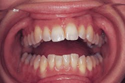
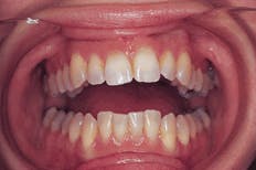
No unusual or abnormal findings were identified during the extraoral examination. Intraoral examination revealed teeth normal in shape and color (see photo). Examination of the oral soft tissues revealed no unusual findings and no bony abnormalities. Radiographic examination revealed teeth with short W-shaped roots and obliterated pulp chambers (see radiograph).
Clinical diagnosis
Based on the information presented, which one of the following is the most likely clinical diagnosis?
- regional odontodysplasia
- dentin dysplasia
- amelogenesis imperfecta
- dentinogenesis imperfecta
- internal resorption
Diagnosis
dentin dysplasia
Discussion
Dentin dysplasia is an autosomal-dominant, inherited disorder characterized by abnormal dentin formation and abnormal pulpal morphology. This rare hereditary condition has been classified into two groups: Type I and Type II. Dentin dysplasia abnormalities are limited only to dentin formation and are not associated with any systemic problems or illnesses.
Clinical and radiographic features
Dentin dysplasia Type I (DDI), or radicular dentin dysplasia, affects the roots and pulp chambers of the teeth; all teeth in both dentitions are affected. Teeth affected by DDI are often referred to as "rootless teeth" because of the lack of root length that occurs due to the defective dentin formation. The radicular dentin undergoes a disorganization during development that results in the dramatic shortening of the roots. If the dentin disorganization occurs early in development, roots may be completely absent or markedly deficient. If the dentin disorganization occurs late in development, only minimal root malformation occurs. The shorter the roots, the more likely the teeth are to exhibit mobility and exfoliate prematurely. Loose teeth are characteristic for this type of dentin dysplasia.
The primary and permanent teeth affected by DDI appear normal clinically; however, radiographic examination reveals defective root formation. Radiographically, the roots of the teeth appear short and blunted or, in some
continued on page 68
continued from page 17
cases, completely absent. The affected roots of mandibular molars are said to have a characteristic W-shaped appearance. The pulp chambers may be completely obliterated or appear as thin crescent or chevron-shaped remnants.
Teeth affected by DDI often have associated periapical radiolucencies in the absence of caries involvement or other obvious causes. Multiple periapical radiolucencies are characteristic for this type of dentin dysplasia.
Dentin dysplasia Type II (DDII), or coronal dentin dysplasia, affects the crowns and pulp chambers of the teeth; all teeth in both dentitions are affected. Only the clinical appearance of the deciduous crowns is affected; the crowns appear bluish-gray or brownish in color and resemble the teeth affected by dentinogenesis imperfecta. In contrast, the crowns of the permanent teeth exhibit no discoloration and have a normal clinical appearance.
Unlike DDI, the roots of the deciduous and permanent dentitions affected by DDII are normal in both shape and length when viewed on a radiograph. The deciduous teeth exhibit obliterated pulp chambers and canals similar to those seen with DDI and dentinogenesis imperfecta. The permanent teeth exhibit pulp chambers that resemble thistle tubes and appear flame-shaped with very narrow pulp canals. Pulp stones (pulpal calcifications) often are visible within the abnormal pulp chambers.
Diagnosis and prognosis
Both DDI and DDII are diagnosed based on clinical and radiographic features. The prognosis for teeth with DDI varies with the severity of the disorder.
The primary treatment concern for DDI is the prevention of tooth loss. Because of the short root formation in DDI, early tooth loss from periodontal disease is common. Therefore, patients must establish and maintain meticulous oral hygiene.
If the roots are extremely short, early tooth loss is almost inevitable and prosthetic replacements are required. If the roots are relatively normal, tooth loss may not be a problem.
The prognosis for permanent teeth affected by DDII is essentially the same as for normal teeth.
Joen Iannucci Haring, DDS, MS, is an associate professor of clinical dentistry, Section of Primary Care, The Ohio State University College of Dentistry.

