A 23-year-old female visited a dentist for evaluation of a palatal swelling. Radiographic
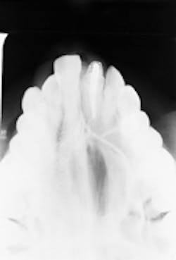
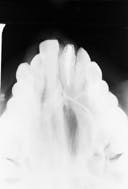
A 23-year-old female visited a dentist for evaluation of a palatal swelling. Radiographic examination revealed a large lesion in the maxilla.
Joen Iannucci Haring, DDS, MS
History
The patient stated that the palatal swelling started several months earlier and recently had become painful. The patient appeared to be in a general good state of health, with no significant medical history. The patient`s dental history included regular dental examinations and routine dental treatment. At the time of the dental appointment, the patient was not taking medications of any kind.
Examinations
The patient`s vital signs were all found to be within normal limits. Examination of the head and neck region revealed no enlarged or palpable lymph nodes. Intraoral examination revealed a bony, hard swelling on the palate measuring approximately 1.5cm x 2.0cm (see photo). No other abnormalities were noted.
The patient`s most recent radiographs were taken approximately one year earlier; no lesion was evident on the previous radiographs. After a thorough clinical examination, a maxillary occlusal radiograph of the involved area was ordered. Examination of the occlusal radiograph revealed a large, well-defined radiolucency (see radiograph).
Clinical diagnosis
Based on the clinical and radiographic examination available, which one of the following is the most likely clinical diagnosis?
- aneurysmal bone cyst
- nasopalatine duct cyst
- simple bone cyst
- central giant cell granuloma
- odontogenic keratocyst
Diagnosis
aneurysmal bone cyst
Discussion
The aneurysmal bone cyst (ABC) is a benign lesion that most often occurs in the shaft of a long bone or in the vertebral column. The cause of the ABC is poorly understood. Some investigators believe it arises de novo, while others claim it represents a reactive lesion - some form of a vascular accident that occurs in a pre-existing lesion. The ABC is considered to be an uncommon lesion of the jaws.
Clinical features
The ABC is most often seen in patients under the age of 20; females are affected more frequently than males. The lesion is most often found in the posterior regions of the jaws, and the mandible is affected more frequently than the maxilla. The ABC is a fast-growing, solitary lesion. Pain is a common feature. Bony expansion is typically present and perforation may occur. The larger lesions are often associated with facial swelling.
Radiographic features
The ABC appears as a unilocular or multilocular radiolucent lesion with marked cortical expansion and thinning. The size of the ABC is variable.
The ABC cannot be diagnosed from its radiographic appearance alone. All lesions identified on a dental radiograph must be documented in the patient record and described in terms of appearance, location, and size. Biopsy and surgical removal must be recommended to the patient.
Diagnosis
A biopsy of the lesion is necessary in order to make a definitive diagnosis. Histologically, the ABC exhibits large, blood-filled spaces surrounded by fibroblastic connective tissue containing giant cells. No epithelial lining is seen; therefore, by definition, the ABC is not a true cyst. Other lesions that may be considered in the differential diagnosis for the ABC include the odontogenic keratocyst, central giant cell granuloma, and ameloblastic fibroma.
Treatment
The ABC is typically treated by curettage. There appears to be a substantial risk for recurrence following treatment; approximately 20 to 50 percent recur after removal. The long-term prognosis is favorable, although more extensive surgical resections may be required to control the recurrent lesions.
Joen Iannucci Haring, DDS, MS, is an associate professor of clinical dentistry, Section of Primary Care, The Ohio State University College of Dentistry.

