Case #9
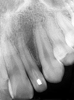
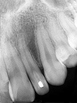
A 27-year-old male visited a dental office for a routine checkup. Radiographic examination revealed an unusual enamel-like radiopacity within the left maxillary lateral incisor.
History
The patient denied any history of symptoms or pain in the areas of the lateral incisor. The patient appeared to be in a general good state of health with no significant medical history. His dental history included regular checkups and routine restorative dental treatment.
Examinations
The patient's vital signs were all found to be within normal limits. Extraoral examination of the head and neck region revealed no enlarged or palpable lymph nodes. Intraoral examination revealed no abnormalities present.
Based on the clinical examination of the patient, selected periapical radiographs, bite wings, and a panoramic film were ordered.
A review of the periapical films revealed a small radiopacity within the crown of the left maxillary lateral incisor (see radiograph). Clinically, the lateral incisor appeared small and peg-shaped. No other abnormalities were noted on the radiographs.
Clinical diagnosis
Based on the clinical and radiographic information available, which one of the following is the most likely diagnosis?
- dens evaginatus
- dens invaginatus
- taurodont
- mesiodens
- regional odontodysplasia
Diagnosis
- dens invaginatus
Discussion
The dens invaginatus (also known as dens-in-dente) is a developmental anomaly characterized by the in-folding (invagination) of the outer enamel tooth surface into the normal tooth structure.
As a result, an enamel-lined structure is seen within the crown (termed coronal dens invaginatus) or root (termed radicular dens invaginatus) of the affected tooth, giving the appearance of a tooth-like structure within a tooth. The term dens-in-dente literally means "tooth within a tooth." The cause of this abnormality is unknown.
Clinical features
The dens invaginatus is most often seen in the permanent lateral incisors, central incisors, premolars, canines, and molars. The maxillary teeth are affected more often than the mandibular. Clinically, the dens invaginatus may be seen as a tooth with a prominent cingulum and a well-defined lingual pit. The pit region is connected to the defect found within the tooth by a narrow constriction. The constriction provides the actual communication from the outside of the tooth to the inner defect.
This configuration is favorable for caries and pulpal necrosis; consequently, the dens invaginatus is associated with a high incidence of pulpal death and periapical disease. In cases where the invagination is extensive, the crown of the tooth appears malformed. Most teeth that are affected by the dens invaginatus do not exhibit changes in crown morphology.
Radiographic features
The dens invaginatus is typically discovered during routine radiographic examination; it has a characteristic radiographic appearance and is readily identified on dental radiographs. The formation of the dens invaginatus takes place early in development and may be viewed on dental radiographs prior to the eruption of the affected tooth.
A range of radiographic appearances may be identified depending upon the extent and severity of this defect. The dens invaginatus may be seen as a coronal invagination or one that involves the root as well. Mild cases may not be apparent on dental radiographs. In severe cases where the crown is malformed, the apical foramen appears wide and abnormally large.
The dens invaginatus typically appears as a pear-shaped invagination of enamel and dentin within the crown and/or root of the tooth that closely approximates the pulp cavity. This in-folding of enamel can be easily recognized on a dental radiograph by its increased radiopacity - it resembles the radiodensity of enamel seen on the external surface of the tooth. Radiographically, the dens invaginatus appears as a small tooth-like structure within the affected tooth. The dens invaginatus is often associated with a periapical radiolucency because of the high incidence of pulpal death and necrosis.
Diagnosis and treatment
The diagnosis of a dens invaginatus is made based on its radiographic appearance. The outline of an enamel-lined invagination within the crown and/or root of a tooth is characteristic. Although each case must be evaluated on an individual basis and treated accordingly, the placement of a restoration in the pit-like defect is the treatment of choice. If the dens invaginatus can be identified on a dental radiograph prior to eruption, plans can be made to place the restoration soon after eruption. The placement of such a restoration should ideally prevent caries, pulpal infection, and premature loss of the affected tooth.
If the pit-like defect is not treated, pulpal necrosis results. Treatment for nonvital teeth affected by dens invaginatus is either extraction or endodontic therapy. The more extensive forms of dens invaginatus may require more sophisticated endodontic care.
Joen Iannucci Haring, DDS, MS, is a professor of clinical dentistry, Section of Primary Care, The Ohio State University College of Dentistry.

