Case #8
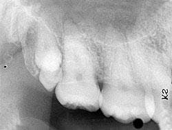
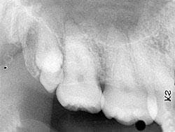
A 25-year-old female visited a dental office for a routine checkup. Radiographic examination revealed two tooth-like radiopacities in the right maxillary tuberosity area.
History
The patient denied any history of symptoms or pain in the right maxilla. The patient appeared to be in a general good state of health with no significant medical history. Her dental history included routine checkups and restorative dental treatment.
Examinations
The patient's vital signs were all found to be within normal limits. Extraoral examination of the head and neck region revealed no enlarged or palpable lymph nodes. Intra-oral examination revealed no abnormalities present.
Based on the clinical examination of the patient, selected periapical radiographs and bitewing films were ordered. A review of the periapical films revealed two tooth-like radiopacities in the right maxillary tuberosity area (see radiograph). The location of the radiopacities was distal to the erupted third molar. No other abnormalities were noted on the radiographs.
Clinical diagnosis
Based on the clinical and radiographic information available, which one of the following is the most likely diagnosis?
- compound odontoma
- complex odontoma
- distomolars
- mesiodens
- paramolars
Diagnosis
- distomolars
Discussion
The distomolar (also known as fourth molar or distodens) is a type of supernumerary tooth. The term supernumerary means in excess of the normal number. Supernumerary teeth, also referred to as hyperdontia, are estimated to occur in 1 to 3 percent of the population. Supernumerary teeth may be seen in both the primary and permanent dentitions. The formation of such extra teeth is a developmental alteration with a strong genetic influence. Many hereditary syndromes are associated with hyperdontia.
Clinical features
Most supernumerary teeth develop within the first two decades of life. Males are affected more frequently than females (2:1). Supernumerary teeth may occur singly or in multiples. A single supernumerary tooth occurs most often in the anterior maxilla while multiple supernumerary teeth are most often found in the mandible. Supernumerary teeth may occur unilaterally or bilaterally. Supernumerary teeth frequently appear smaller than normal-sized teeth. The shape of a supernumerary tooth may vary from normal to conical. Although supernumerary teeth may erupt or remain impacted, most do not erupt due to the lack of available space.
Supernumerary teeth may be found in any location in the jaws. The two areas most frequently involved include the maxillary midline area and the areas distal to the third molar areas of the jaws. Following these most common sites of occurrence, the other regions that may exhibit supernumerary teeth include the premolar, canine, and lateral incisor areas. There are specific terms used to describe supernumerary teeth based on location: mesiodens, distomolar, and paramolar.
The mesiodens is a tooth (-dens) found near the midline (mesio-) of the maxilla. It is the most common supernumerary tooth. The mesiodens is a small tooth with a conical-shaped crown and a short root. Approximately 75 percent of mesiodens fail to erupt. When impacted, the mesiodens most often presents in an inverted position.
The distomolar, as the name implies, is located distal to the third molar. It is the second most common supernumerary tooth. This tooth does not interfere with the normal eruption pattern of the first and second molars. The distomolar may resemble a normal molar in size and shape, or may appear rudimentary and miniature. The distomolar may erupt or remain impacted.
The paramolar is a supernumerary tooth that occurs around (para-) the molar region in a buccal or lingual orientation. As with the distomolar, this tooth may appear normal in shape and size, or rudimentary and miniature.
Radiographic features
Dental radiographs (periapical, bitewing, and panoramic films) can be used to identify unerupted supernumerary teeth. Such teeth can be identified on a radiograph after the ages three or four (primary dentition) and after the ages nine to 12 (permanent dentition). Although the shape and size of supernumerary teeth may vary, the radiographic appearance of the tooth (radiopaque enamel and dentin) is identical to the other teeth on the film.
Diagnosis and treatment
The diagnosis of a supernumerary tooth, including the distomolar, is made based upon the characteristic radiographic appearance and/or clinical presentation. Less than 20 percent of supernumerary teeth exist without clinical complication. Non-erupted supernumerary teeth exist without clinical complication. Non-erupted supernumerary teeth may be associated with cyst development, root resorption, or the impediment of tooth eruption. Therefore, the recommended treatment for such impacted teeth is surgical removal. The decision to surgically remove a supernumerary tooth depends upon the location, position and number of teeth, and the potential complications that may occur from the surgical removal.
Erupted supernumerary teeth may impede the eruption of normal teeth and cause crowding or malpositioning of adjacent teeth. Erupted, nonfunctional supernumerary teeth should be extracted. Patients that present with multiple supernumerary teeth should be evaluated for syndromes associated with hyperdontia, such as Gardner syndrome or Cleidocranial dysplasia.
Joen Iannucci Haring, DDS, MS, is a professor of clinical dentistry, Section of Primary Care, The Ohio State University College of Dentistry.

