A 20-year-old male college student visited a dentists office for evaluation of multiple oral
A 20-year-old male college student visited a dentist`s office for evaluation of multiple oral lesions.
Joen Iannucci Haring, DDS, MS
History
The patient stated that the sores in his mouth had started several days earlier. He described the sores as painful. When questioned about constitutional symptoms, the patient claimed an overall feeling of malaise, runny nose, and a low-grade fever. The patient denied any history of a previous episode.
The patient`s medical history was reviewed. No significant findings were noted. At the time of the examination, the patient was taking Tylenol® for the oral pain.
Examinations
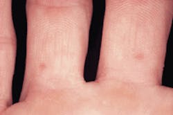
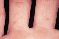
Examination of the head and neck regions revealed enlarged superficial cervical and submandibular lymph nodes. All vital signs were within normal limits. No skin lesions were apparent in the head and neck regions. Examination of the skin on the hands and feet revealed multiple scattered red and vesicular lesions on the hands (see photo). Oral examination revealed multiple ulcerations scattered throughout the oral cavity (see photo).
Differential diagnosis
Based on the clinical information provided, which one of the following is the most likely diagnosis?
* Hoof and mouth disease
* Hand, foot, and mouth disease
* Erythema multiforme
* Primary herpetic gingivostomatitis
* Herpetiform aphthous ulcers
Diagnosis
__ Hand, foot, and mouth disease
Discussion
Hand, foot, and mouth disease is a highly contagious disease caused by the coxsackieviruses. The coxsackieviruses belong to the genus enterovirus in the family of viruses known as Picornaviradae. Enteroviruses were first discovered as a result of research efforts concerning the development of better methods for growing the poliovirus.
In 1948, investigators were searching for suitable animals to replace monkeys in the existing poliomyelitis studies. These researchers inoculated mice with fecal suspensions from two suspected poliomyelitis cases. The mice became paralyzed, not with the poliovirus, but with the first isolate of a new virus group that was subsequently named for the hometown of the patient, Coxsackie, N.Y.
Further studies with mice identified two groups of the coxsackievirus, group A and group B. Many illnesses were soon recognized as being related to these coxsackieviruses. During the next 20 years, numerous and distinct viral isolates were described; the isolates were numbered sequentially within the groups coxsackie A and coxsackie B.
The routes of transmission for the enteroviruses are the same as for the poliovirus. Although enteroviruses are isolated in the highest titers and for the longest period of time in stool specimens, respiratory secretions may also be involved. Therefore, fecal-oral transmission and spread via contact with respiratory droplets are considered to be the most likely modes of transmission.
The enteroviruses gain entrance to their human host via the oropharynx and then replicate in the epithelial and lymphoid cells of the intestinal tract. From there, the viruses spread to other sites via and blood stream. The enteroviruses are shed from the oropharynx and urine for one week, and in the feces for up to one month. The incubation period for enteroviruses is short, usually less than 10 days.
The diseases associated with the enteroviruses tend to occur in epidemics. Outbreaks may occur in small groups (schools or day care centers, for example) or in selected communities. Outbreaks may also become widespread and affect regional, national, or international areas. In temperate climates, most cases occur in the summer or early autumn. In tropical climates, there is no seasonal pattern. Poor hygiene and crowded conditions facilitate the spread of these viruses.
Enterovirus infections can result in a wide variety of disease syndromes. Most often, enterovirus infections are subclinical or present as mild, upper respiratory-tract symptoms. Central nervous system diseases, including aseptic meningitis and encephalitis, are the most commonly recognized serious manifestations of enterovirus infection. A number of enterovirus serotypes have also been associated with severe illness in newborns, including sepsis and general disseminated disease. The two enterovirus diseases that have oral implications include hand, foot, and mouth disease and herpangina.
Hand, foot, and mouth disease (HFM) is caused by the coxsackievirus A16, and to a lesser extent, coxsackieviruses A5, A7, A9, A10, B2 and B5. The first recognized outbreak of this disease was in Toronto in 1957, although the disease was not named as "hand, foot and mouth" until an epidemic in England in 1959. Since then, the disease has occurred worldwide.
Clinical features
Although HFM may occur in either children or adults, most cases arise in children between the ages of six months and five years. Constitutional symptoms include an overall feeling of malaise, cervical lymphadenopathy and low-grade fever. Coryza, or the profuse discharge from the mucous membranes of the nose, may also be present.
Clinically, HFM is characterized by lesions that affect the skin and the mouth. A vesicular skin eruption occurs on the skin of the hands and the feet, and, to a lesser extent on the arms, legs, and buttocks. The cutaneous lesions number from one to 100 and present as short-lived vesicles surrounded by an erythematous halo. The vesicular fluid contains the coxsackievirus and provides a mode of spread through contact. The vesicles rupture and ulcerations result. In less than 15 percent of cases, no skin lesions occur. The skin lesions appear concomitantly or shortly after the oral lesions.
The oral lesions of HFM are characterized by vesicles that rupture and leave painful, raw erosions and ulcerations. The multiple lesions may be found anywhere in the oral cavity. The palate, buccal mucosa, and tongue are the most frequent sites of occurrence. The gingiva and oropharynx may also be involved. A "sore mouth" is often the chief complaint for a patient with HFM. In children, refusal to eat is common.
Differential diagnosis
The oral vesicular-ulcerative lesions of HFM may resemble a number of disease entities. The characteristic distribution of lesions on the hands and feet eliminates other diseases from the differential. In cases where no lesions are seen on the hands and feet, diseases such as primary herpetic gingivostomatitis, erythema multiforme, and herpetiform recurrent aphthous ulcers should be considered in the differential diagnosis.
Diagnosis
HFM is often diagnosed on a clinical basis and is established based on the clinical signs, symptoms, and patient history. In instances of questionable diagnosis, laboratory confirmation may be required. Cytologic smear techniques may be used to diagnose HFM; intracytoplasmic viral inclusions may be demonstrated in vesicular scrapings of the lesions. Diagnosis may also be made via viral culture. However, this test is both slow and expensive.
Treatment
HFM is a self-limiting disease, and recovery usually occurs within one to two weeks. As with many other viral diseases, there is no specific treatment. Supportive therapy includes antipyretics to reduce fever and analgesics to control pain. Palliative rinses may be recommended to alleviate oral discomfort. In small children, viscous lidocaine may be used. HFM is not a recurrent disease. Following infection, a permanent immunity develops.
HFM vs. hoof and mouth disease
HFM should not be confused with hoof and mouth disease. Despite the similarity in names, HFM bears no relationship to hoof and mouth disease. Hoof and mouth disease is a viral infection that affects livestock animals. This disease is seen in hogs, sheep, cattle, and rarely, man. When hoof and mouth disease is seen in humans, it is transmitted via contact with infected animals or animal products.
In humans, hoof and mouth disease is characterized by malaise, fever, nausea, vomiting and vesiculo-ulcerative lesions of the oral mucosa and pharynx. Skin lesions involve the feet and oral cavity. Hoof and mouth disease was common around the turn of the century. Today, hoof and mouth disease is nearly nonexistent in the United States due to stringent cattle importation laws and effective domestic health laws.
Joen Iannucci Haring, DDS, MS, is an associate professor of clinical dentistry, Section of Primary Care, The Ohio State University College of Dentistry.

