Oral pemphigus vulgaris
by Nancy Burkhart, RDH, EdD
The patient that you are treating in your practice today is a male, age 47, whose name is Mr. Wallace. He is married, with two children, and is new to your practice. A review of his health history suggests that he has been diagnosed with pemphigus vulgaris during the past six months. You quickly retrieve all the information from your knowledge bank and try to remember what treatment and precautions are suggested for this type of autoimmune disease. Mr. Wallace has hypertension and currently takes a hydrochlorothiazide medication, and he has been prescribed a low dose of prednisone. No other significant health findings are apparent from his health history.
Notes and findings: Mr. Wallace has recent radiographs that were taken at his former dental office. As you review the radiographs, you notice some posterior molar bone loss, but no other significant findings. Mr Wallace also tells you that he has some skin lesions. The areas that are visible to you appear small and crusted on the extremities. He is being treated by both an internal medicine expert and a dermatologist.
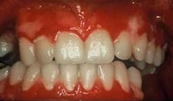
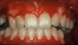
Clinical impressions: As you begin your oral assessment, you note the buccal mucosa and gingiva are erythematous with some ulceration and erosion. The erythema is very prominent at most marginal gingival areas. The tissue bleeds with very little pressure (see figure 1). You also notice that his lips appear ulcerated and crusted (see figure 2).
Diagnosis: Pemphigus vulgaris
Etiology: Pemphigus is a term used to describe a group of chronic skin diseases with potential intraoral involvement including pemphigus vulgaris, pemphigus vegetans, pemphigus foliaceus, and pemphigus erythematosus. Pemphigus vulgaris is by far the most common form of the disease. Patients tend to be affected most often in midlife with Jewish and Mediterranean individuals affected more often than other populations. Pemphigus is classified as an autoimmune disease that forms suprabasilar bullae and the overlying tissue is easily disrupted or stripped.
Method of transmission: Pemphigus is an autoimmune disorder and is not transmitted from one individual to another.
Pathogenesis: Pemphigus may affect any mucous membrane such as the esophagus, larynx, pharynx, vagina, and anus. External skin surfaces may be affected as well. Before the use of corticosteroids in the 1950s, pemphigus vulgaris was almost uniformly fatal within five years, and it continues to be a life-threatening disease. The use of prednisone has reduced mortality and continues to be instrumental in the treatment of pemphigus vulgaris.
Perioral and intraoral characteristics: Pemphigus vulgaris usually affects the skin and oral tissues, but lesions in the mouth may precede skin lesions. The dental team may be the first health-care members to notice the oral lesions that occur in pemphigus vulgaris, and they are a crucial link, assisting the patient in obtaining the necessary medical care that is needed.
Early treatment is crucial in controlling pemphigus vulgaris. Sirois, et al. (2000) reported that more than 50 percent of patients in their study sought initial care from their dental clinician. Just as pemphigoid (March 2007) is often characterized as desquamative gingivitis (a general term), this is true also for pemphigus vulgaris depending upon the severity of the disease. The tissue involved is erythematous and ulcerated (see figure 1). Bullae may be present and they rupture to form painful erosions. The borders of the erosions are often ragged and painful. Depending upon the type of pemphigus, the lesions may appear to have vegetations or scales on the surface.
Extraoral characteristics: Patients with untreated pemphigus vulgaris will develop skin lesions, so any observed skin lesions that are visible should be fully evaluated. Skin lesions are treated with various topical and systemic steroids/corticosteroids and these are the main line of therapy. The medications are increased or decreased depending upon the severity of the lesions.
Several new treatments such as the use of intravenous immunoglobulin, monoclonal antibodies, mycophenolate mofetil (CellCept) are used as well. When skin lesions are extensive, an adjunct type therapy, hyperbaric oxygen therapy (HBO) has been suggested in addition to normally prescribed medications. HBO therapy is reported to have antiseptic and possibly immunosuppressive properties. The theory is that over-oxidization of the keratinocyte membrane seems to decrease its antigenicity (having the properties of an allergen), resulting in an impaired fixation of circulating pathogenic PV autoantibodies. This reduces the immune response characteristics of the disease. It has been found beneficial in treating infections such as osteomyelitis (Lentrodt, et al. 2007) as well.
Distinguishing characteristics: The stripping of the epithelium in pemphigus vulgaris represents a positive Nikolsky’s Sign, and this is a key characteristic of the disease. The oral epithelium can be separated at the basal cell layer and rubbed off when pressed with a sliding motion.
Significant microscopic features: With all vesiculobullous diseases, a histological stained biopsy should be performed. Depending upon the tissue sample, the histological sample is not always conclusive. If the epithelium is intact, the H & E stained tissue sample will present with a suprabasilar bullae formation and perivascular edema. Free-floating epithelial cells (Tzanck cells) are observed (see figure 3). An immunofluorescence stain of the tissue provides a conclusive diagnosis and is usually diagnostic because of the distinct pattern of the stain (see figure 4). The adhesions of the intracellular material that bind the cells together are affected. Acantholysis is the term used to describe this cellular separation. As seen in figure 4, each cell is very distinct when immunofluorescence is performed.
Differential diagnosis: Other vesiculobullous diseases must be considered including lichen planus, pemphigoid, lupus, epidermolysis bullosa acquisita and erythema multiforme. Clinically, the tissue may have very similar appearances exhibiting raw, ulcerated, erythematous tissue.
Treatment and progress: Pemphigus vulgaris causes ulcerated areas that are painful for the patient. Patients may limit their diets to soft foods for the sake of comfort, but this often leads to more plaque accumulation. Meanwhile, patients cannot brush effectively due to pain. This may lead to an increased incidence and severity of dental caries and periodontal disease. When restorative work is performed such as multiple crowns, implants or partial/full dentures, the tissue becomes extremely problematic with extensive ulcerations.
Maintenance appointments with the hygienist need to be conducted in a gentle fashion with multiple appointments for debridement in severe cases. Ideally, there should be as little disruption to the tissue as possible. Often, using simple hand scaling instruments is most effective and allows the hygienist more control in tissue manipulation of patients who have severe mucosal disease. Simply polishing with a nonabrasive toothpaste is all that is needed.
As with other mucosal disease states such as MMP and OLP, gentle tissue manipulation is the best course of treatment for the patient. The use of air polishers and harsh abrasives should be avoided since the tissue is so fragile. Additionally, more frequent maintenance appointments for debridement are recommended with meticulous plaque control. Alcohol, spicy foods that burn, and harsh mouth rinses containing alcohol are contraindicated in the treatment of pemphigus vulgaris.
Often, short-term, high-dose prednisone is necessary and is often the main line of therapy. After a period of time, the dosage can be adjusted for the individual patient when the lesions are under control. Potent topical steroids are used to control oral pemphigus vulgaris, just as these are used to treat and control other mucosal diseases such as lichen planus, mucous membrane pemphigoid, and lupus.
The individual patient and the extent of the ulceration are always considerations. According to Ruocco, et al. (2000), “The goals of therapy are to suppress blistering, control progression of the disease, facilitate and optimize re-epithelialization, prevent complications or sequelae, and cope with the side effects of the drugs (or other treatments) employed.” Both medical and dental teams are needed for treatment of the patient with oral pemphigus vulgaris. It should be emphasized that they need to engage in dialogue to convey the best treatment for the patient.
With the long-term use of both systemic and topical steroids, patients may develop candidiasis infections and may need treatment periodically for candida. Often antifungal type medications are prescribed in the form of rinses, troches, and ointments such as Nystatin and Diflucan, etc. The problem with candida is usually an ongoing one with the practitioner working back and forth to balance the steroid use with the development/treatment of the candida infections. The hygienist should be alerted to the typical oral implications and identification of candida in patients with pemphigus vulgaris.
Nancy Burkhart RDH, EdD, is an adjunct associate professor in the Department of Periodontics at Baylor College of Dentistry, Texas A & M Health Science Center in Dallas. Nancy is also a co-host of the International Oral Lichen Planus Support Group through Baylor (www.bcd.tamhsc.edu/lichen). She can be contacted at [email protected].
References:
Endo H, Rees TD, Matsue M, Kuyama K, Nakadai M, Yamamoto H. Early detection and successful management of oral pemphigus vulgaris: a case report. J Periodontol. 2005 Jan; 76(1):154-60.
Fatahzadeh M, Radfar L, Sirois DA. Dental care of patients with autoimmune vesiculobullous diseases: Case reports and literature review. Quintessence Int 2006:37:777-787.
Lentrodt S, Lentrodt J, Kubler N, Modder U. Hyperbaric oxygen therapy for chronically recurrent mandibular osteomyelitis in childhood and adolescence. J Oral Maxillofac Surg. 2007 Feb; 65(2):186-91.
Plemons JM, Gonzales TS, Burkhart NW. Vesiculobullous diseases of the oral cavity. Periodontology 2000, 1999:21; 158-175.
Ruocco V, Ruocco E, Wolf R. Bullous Diseases: Unapproved treatments or indications. Clinics in dermatology 2000;18:191-195.
Sirois DA, Fatahzadeh M, Roth R, Ettlin D. Diagnostic patterns and delays in pemphigus vulgaris: Experience with 99 patients. Arch Dermatol 2000; 136:1569-1570.

