Aphthous ulcers
by Nancy Burkhart, RDH, EdD
Presentation: Mr. Patel, a 40-year-old male, is your patient today. Mr. Patel was last seen six months ago for a maintenance appointment. He has no medical or dental issues, and he considers himself to be in very good health. His only complaint is the lesion on the labial mucosa that he has noticed for the past two days.
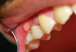
Notes and findings: Upon examination, you notice a lesion approximately 2-3 millimeters in diameter with a yellow-white center and a red outer rim. The patient reports some sensitivity, but he does not consider the lesion to be painful. The patient also reports that he has not had a fever in the past few days (see Figure 1).
Clinical impressions: The area in question is clearly ulcer-like, but the patient does not report pain. He does, however, say that it was initially uncomfortable. Mr. Patel tells you that he gets ulcers periodically and mostly on the ventral surface of the tongue. He remembers trying a new Waterpik instrument and scraping the mucosal area with the tip of the device.
Diagnosis: Aphthous ulcer. The ulcers are also called recurrent aphthous ulcer, canker sore, aphthous stomatitis, or Mikulicz ulcers. Aphthous ulcers are sometimes confused with traumatic ulcers and herpetic lesions.
Etiology: Aphthous ulcers occur in approximately 20 percent of the population in the United States and up to 66 percent of those who have aphthae are adolescents or young adults.3 Aphthous ulcers are commonly seen in a dental practice, and the hygienist will most likely have several patients who suffer from recurrent aphthae. The etiology is not clearly understood. But they have been associated with trauma, dietary sources and food allergies (celiac sprue), immunology, menstrual cycles, sun exposure, genetic predisposition, vitamin deficiency, sodium lauryl sulfate, smoking cessation, and stress-related factors.
In the case that has been presented, trauma to the mucosa with the instrument tip of the oral irrigator device is a suspected contributor to the ulcer development in this susceptible individual. Patients who are prone to this type of ulcer may traumatize the tissue by merely eating rough foods or drinking hot liquids.
Method of transmission: The literature suggests that the aphthous ulcer cannot be transmitted to another individual.
Pathogenesis: There may be an immunological component in some patients with recurrent aphthous ulcers,5 and members within the same family may be more prone to the development of these ulcers. Hypersensitivity reactions, allergens, gastrointestinal disorders, vitamin deficiencies, endocrine factors, immune deficiency, and anemia have been implicated in recurrent aphthae. A full hematologic screening can be beneficial in recurrent cases.6
Interestingly, patients who use tobacco products may be somewhat protected from aphthous ulcer development. The cause of this is not known, but it has been suggested that smoking may increase the keratinization of the oral tissues either due to heat or the direct effect of tobacco chemicals. Additionally, a substance in nicotine may play a role in making the individual less susceptible, since some reports suggest that patients on nicotine replacement enjoy the same type of immunity. This should be kept in mind with regard to patients who are going through smoking cessation or those who have recently quit using tobacco products.
Three types of ulcer formation have been described:
• RAU minor - Minor aphthae, also called Mikulicz ulcer, are far more common that the other types. The ulcer usually measures approximately 2-4 millimeters in diameter and always less than one centimeter. The age group is in the range of 20 to 40 years of age, and the lesions lasts approximately seven to 10 days. The herpetic type ulcer, caused by the herpes simplex virus, is preceded by vesicles that rupture.
by the herpes simplex virus, is preceded by vesicles that rupture.
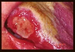
• RAU major - The major type is much larger and measures from 1-3 centimeters. Approximately one in 10 aphthae sufferers has the more severe form.3 These lesions are deeper and more crater-like with a longer duration and healing time. The major form is sometimes called Sutton’s disease or periadenitis mucosa necrotica recurrens, and scarring of the tissue is reported in most cases (see Figure 2).
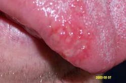
• RAU herpetiform - The herpetiform type measures from 1-3 millimeters in diameter, tends to be more numerous (10 to 100 at any given time), and collects in clusters (see Figure 3). The lesions occur on both keratinized and non-keratinized tissue and may be confused with herpetic gingivostomatitis. Although all aphthae are surprisingly painful in some cases, the herpetiform type may be particularly uncomfortable. The lesions may take 10 days or longer to heal and they tend to recur rather frequently. This form is found in a slightly older age group and has a higher predilection for females. RAU herpetiform also begins with vesicles that rapidly develop into pinhead-sized ulcers. This sometimes adds to the confusion with herpetic gingivostomatitis. However, the HSV cannot be cultured from these lesions since the herpes virus does not cause them. A cytology smear would confirm a diagnosis.
Perioral and intraoral characteristics: Most aphthae present as single lesions with a yellow center and a red, erythremic border as depicted in Figure 1.
Extraoral characteristics: Aphthous ulcers may occur on the labial mucosa and occasionally extend onto the vermilion border of the lip. Patients often have a tingling, burning, or slight tightening sensation prior to the development of the actual ulcer (this is termed prodromal sensation). Confusion occurs sometimes with the herpetic type lesions (herpes simplex virus) that are seen both intraorally and periorally as herpes labialis on the lips and commissures.
Distinguishing characteristics: Aphthous ulcers are usually found on movable, non-keratinized mucosa and present as shallow ulcers with a definite clinical appearance of an erythematous “halo” and a yellowish center. Oral non-keratinized mucosa would be the buccal and labial mucosa, the vestibule, the ventral surface of the tongue, the floor of the mouth, the soft palate, and oral pharyngeal areas. As stated previously, the herpetiform aphthous variety and herpetic gingivostomatitis (HSV) may appear in any area of the mouth.
Significant microscopic features: The microscopic features are usually not diagnostic since the presence of lymphocytes, macrophages, and mast cells are common to many other inflammatory lesions. The epithelium is usually disrupted or absent, with the lesion extending into the lamina propria, as would be expected in a cratered lesion.
Differential diagnosis: The most difficult problem in diagnosis for a practitioner would be ulcers that do not appear clearly aphthous in nature. The ulcer in the slide of the patient presented here fits all the diagnostic features of the aphthous ulcer with regard to location, color, size, and history. The presence of large ulcers and the frequency of the ulcers may make the clinician curious about other systemic diseases that may be an underlying factor.
Considerations may be: hand, foot, and mouth disease; AIDS; Reiter’s syndrome; Behcet syndrome; celiac sprue; vitamin deficiencies; Crohn’s disease; systemic lupus; and PFAPA syndrome.4 Ulcers can be associated with other disease states such as syphilis, tuberculosis, blood dyscrasias, and dermatoses.1 With any undiagnosed lesion, cancer is always a consideration until proven otherwise. Some of these disease states will be discussed in future oral case studies.
Treatment and prognosis: Aphthous ulcers usually clear within a week regardless of treatment. Some patients may report discomfort while others will have no pain and will not need any type of pain medication. Treatments with rinses of chlorhexidine gluconate and topical corticosteroids are sometimes prescribed when needed. Topical ointments such as Kenalog in Orabase may be used as well, but warm saline rinses are usually all that is needed in mild cases.
New products on the market include a small, round, covered bandage that fits over the ulcer and becomes a gel-like patch when placed in the mouth. The cover may protect the ulcer from further injury from trama and spicy or hot foods. Additionally, other gels may be herbal based with aloe vera, lysine, and echinacea that coat the ulcer and claim to aid in the healing process.
Encouraging the patient to keep a diary so that patterns may be established can be very beneficial. Reduction of known trigger mechanisms such as stress, trauma, sunlight, and trigger foods is suggested. A diet that may eliminate trigger mechanisms is helpful in preventing future recurrences. Limiting spicy or citrus foods is often helpful in reducing discomfort. Elimination of known dental products that may contribute to future aphthae is warranted since some patients may be sensitive to sodium laurel sulfate and flavoring agents. Minimal disruption of the oral tissues during oral hygiene procedures is a consideration as well.
Nancy Burkhart, RDH, EdD, is an adjunct associate professor in the Department of Periodontics at Baylor College of Dentistry, Texas A & M Health Science Center in Dallas. Nancy is also a co-host of the International Oral Lichen Planus Support Group through Baylor (www.bcd.tamhsc.edu/lichen). She can be contacted at [email protected].
References
1 Gill Y, Scully C. Mouth ulcers: a study of where members of the general public might seek advice. British Dental Journal Published online: 2 February 2007/doi:10.1038/bdj.2007.82
2 River-Hidalgo FR, Shulman JD, Beach MM. The association of tobacco and other factors with recurrent aphthous stomatitis in a US adult population. Oral Diseases 2004: 10, 335-345.
3 Porter SR, Scully C, Pedersen A. Recurrent aphthous stomatitis. Crit Rev Oral Biol Med. 2003; 9:1499-1505.
4 Rees TD, Binnie WH. Recurrent aphthous stomatitis. Dermatologic Clinics. 1996; April. 14:2, 243-56.
5 Scully C, Felix DH. Oral medicine: update for the dental practitioner. Aphthous and other common ulcers. British Dental Journal 2005;199: 259-264.
6 Ship JA, Chavex EM, Doerr PA, Henson BS, Sarmadi M. Recurrent aphthous stomatitis. Quintessence Int. 2000; 31:95-112.


