Soft Tissue Recontouring With The Diode Laser
by David P. Reichwage, DDS, and Kim Lemberg
All ceramic bonded restorations using contemporary materials and techniques for enhancement of esthetics of anterior teeth are very predictable in terms of function and appearance. The long-term success of bonded restorations is dependent on maximizing the health and integrity of the gingival tissues. The gingival symmetry that embraces the elements of smile design requires optimal soft-tissue health at the free gingival margin, absence of bleeding on probing or scaling, attached gingiva that is stippled and of normal color, and sulcus pocket depths of 1-3 mm.
These same principles apply to the natural dentition as well. They are guidelines to construct a "blueprint" to maximize soft tissue esthetics where harmony is created with the upper and lower lips, tooth position, size, and shape. These principles include golden proportion of maxillary anterior dentition, midline and arch alignment, axial inclination, incisal edge and lower lip alignment, contact points of maxillary anterior dentition, arch form, graduation of teeth posteriorly, gingival symmetry, and gingival contour.
History
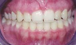
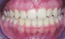
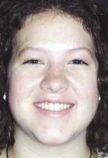
A 20-year-old female presented with the following concerns. She had finished orthodontics about four years ago. She felt tooth position and arch form were favorable; however, the soft tissue height and contour did not complement her teeth (see Figure 1). She thought her teeth were too short and square and that the gum tissue appeared blunted and irregular. In addition, her maxillary frenum was very low and attached to the papilla between teeth No. 8 and No. 9. She was planning her wedding and wanted a "new and better smile" (see Figure 2).
Procedure
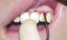
The objectives and goals for soft-tissue correction were presented to the patient and her parents. Periapical radiographs, periodontal charting, and models were used to develop the treatment plan. Radiographs showed excellent bone level, and pocket depths ranged between 3.0 to 3.5 mm. The width of teeth No. 8 and No. 9 was 8 mm and length was 8.5 mm. A marker was used on the soft tissue to simulate the amount of tissue reduction required to produce the length of teeth Nos. 6 through 11 in proportion to the width (see Figure 3).
The golden proportion refers to the visual width of the maxillary anterior teeth. Measurements for the lateral incisors are divided into the measurements for centrals and cuspids. Ideally, the centrals relative to the lateral is 1.618 and to the cuspids 0.618. The midline is the axis running down the center of the face. The arch alignment should be perpendicular with the midline or parallel with the eyes. The axial inclination of the maxillary teeth has a vertical axis, which is inclined slightly to the mesial. The axis is a line drawn from the gingival apex to the center of the incisal edge.
In a natural smile, the incisal edges of the maxillary dentition should follow the contour of, but not contact, the lower lip. The interproximal contact points of the central incisors should be in close proximity to the incisal edge. They gradually progress gingivally as they move posteriorly. The arch form of the maxillary teeth is determined by a line drawn between the tip of the canines — bisecting the incisive papilla. The graduation of the posterior teeth should be geometrically symmetrical. The teeth should appear shorter descending from canines to molars.
When the gingival tissue is exposed during a wide smile, it is important that the gingival height of corresponding teeth on each side be symmetrical. This symmetry is critical as the midline is approached because the close proximity makes the factor obvious. The gingival apex of the lateral incisors is lower than that of centrals and canines. This apex is 1-2 mm below a line drawn between the apex of the centrals and canines. The gingival zenith is the highest point of contour at the apex and should be just distal to the long axis of central and canines — but at the center for the laterals. This was determined to be 0.75-1.5 mm from study models analysis without violating the biological zone of attached tissue.
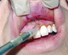
Emmela topical anesthetic was applied on the tissue from teeth Nos. 5-12 and lidocaine HCl 2% with epinephrine was administered with a 30-gauge needle.
The diode laser with Touch Tips was used for tissue reduction (see Figure 4). The diode laser provides quick, very precise soft-tissue surgeries with minimal or no bleeding, swelling, or postoperative pain. Hemostasis is achieved and edema is prevented because capillaries and lymphatics are cauterized and sealed. In addition, tissue damage from the procedure is minimal. Since tissue damage is related to the total energy delivered over time, the Touch Tips and fiber are used in continuous, sweeping strokes and are kept moving during lasing.
A cervical shaped Touch Tip was used with the diode laser on a setting of 3.5 watts continuous wave. The Touch Tip was angled toward the incisal and gingiva was contoured to the desired dimensions using a constantly moving stroke. A chisel-shaped Touch Tip was then used to bevel the gingival margin.
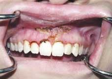
The maxillary frenum to be released was drawn tight by retracting the lip. A conical Touch Tip was used with the diode laser set at 4.0 watts continuous wave to make a releasing incision at the base where the frenum attaches to the maxilla. The muscle attachment was followed laterally and apically to minimize reattachment (see Figure 5). The completed surgical area had the classic diamond shape (see Figure 6). Since traditional scalpel incisions were not used, sutures were not needed.
Post-operative care directions given to the patient were doxycycline 100 mg BID for one week, Dolobid 500 mg, and Oxygel to be applied to the affected area two times a day. A post-op check was done the following day and the patient reported that she experienced "slight discomfort." On a scale of 1-10, the patient rated her pain as a "two." This lasted one day, during which she took Dolobid. For approximately one week following, she reported having to take only Tylenol as needed.
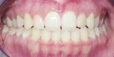
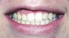
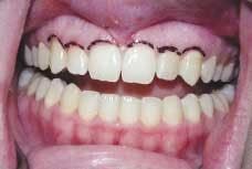
The gingival contouring healed in four to five days (see Figure 7). The patient was very pleased with the results of the procedure and reported that her friend said, "It is beautiful! It looks like it has been airbrushed, like in a magazine. It is so perfect!" (see Figure 8)
Conclusion
The diode laser represents the latest advance in laser technology. It is a surgical instrument that is reliable, portable, and powerful. It provides quick, precise soft tissue surgeries with minimal or no bleeding, swelling, or post-operative pain. It has a broad range of applications, which include tissue retraction during restorative procedures, gingivectomy, gingivoplasty, crown lengthening, frenectomy, bacterial decontamination, and removal of diseased epithelial lining during periodontal treatments. Its use enhances placement of contemporary materials and enables soft tissue corrections both for restorations and with the natural dentition.
David P. Reichwage, DDS, maintains a private practice in Fort Wayne, Ind. He holds program certifications from the Las Vegas Institute for Advanced Dental Studies, Pacific Esthetic Continuum, Academy of Laser Dentistry, JP Consultants Institute, Comprehensive Care Consulting, and Dr. James Carlson. Dr. Reichwage is a member of the Academy of Operative Dentistry, Academy of Laser Dentistry, ADA, Indiana Dental Association, and Isaac Knapp District Dental Society. He has served organized dentistry on both state and local levels and is a past president of the Isaac Knapp District. Kim Lemberg is the hygiene coordinator/concierge at Fort Wayne Smiles, Dr. Reichwage's office. Kim also holds certification from The Las Vegas Institute for Advanced Dental Studies.