Oral pathology: Noting a pigmented macule that often presents a diagnostic dilemma
By Nancy W. Burkhart, BSDH, EdD
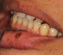
Your patient today is Ling, a 43-year-old Chinese-American female who is a new patient in your practice. As the extraoral exam begins, you notice a pigmented macule on her lower lip. Upon questioning her, she states that the pigmentation has been there for as long as she can remember. She reports that there has never been an issue with the pigmented area such as irritation, bleeding, or ulceration.
Ling wants to know why she has this darker pigmentation and if there is any way to remove it.
A focal pigmented lesion that occurs on the lip or the intraoral tissues is referred to as an oral melanotic macule or labial melanotic macule when occurring on the lip. The oral melanotic macule is the most common oral lesion of melanocytic origin. There is an increase due to melanin deposits in the basal cell layer, lamina propria, or both in the microscopic characteristics of the macule.
Labial melanotic macules (occurring on the lips) most often are seen in adults. But as stated in Ling’s case history, they may be present early in life. The macules appear as single lesions that are pigmented, flat, and well delineated. They are usually uniform in color and sun exposure is not an etiology. The intraoral lesions usually occur in patients older than 40. However, they may occur at any age and affect approximately 3% of the population, with the lip being the most common location followed by the gingiva. Focal melanosis is more commonly found in females.
The melanosis may occur on the vermilion border of the lip or the gingiva due to postinflammatory or posttraumatic causes. Melanotic macules usually arise from three sources: an intraoral freckle, postinflammatory pigmentation, or disorders such as Addison’s disease, Peutz-Jeghers syndrome, or Laugier-Hunziker syndrome. Although the melanotic macule does not develop due to sun exposure, its origin is not conclusive. Inflammation is known to promote pigmentation such as in smoker’s melanosis and chronic oral inflammation from disease states.
These macules are most often seen on the lip, followed by the palatal, gingival, and buccal mucosa. Melanotic macules that occur on the buccal mucosa are usually multiple. A differentiation is usually made when deciding whether the pigmentation is an ephelis (freckle) or a melanotic macule. When occurring on the lip, the appearance of a freckle or ephelis tends to be less than 0.5 cm, and they are associated with sun exposure. This is in contrast to the focal melanotic macule, which is not associated with solar damage and tends to be larger. Ephelides (plural of “ephelis”) are usually found in fair-skinned individuals, are due to sunlight exposure, occur in childhood, tend to diminish with age, and are lighter in appearance. The melanotic macules are usually brown, deep blue/black, or black, and they are well circumscribed with sharp, delineated borders.
Ephelides often may be indistinguishable from melanotic macules without viewing them under the microscope. Histologically, ephelides have fewer melanocytes but have more melanosomes, which are larger in size. Using a sunscreen of 15 SPF will help to fade the ephelis and discourage new ones from forming.
In contrast, solar lentigines are usually seen in older individuals, are much larger than ephelides, and tend to persist indefinitely even with sun protection. The macules are larger and appear more pigmented than ephelides. A good example would be the backs of the hands of a fair individual who has been exposed to the sun over many years.
When changes occur with these lesions, such as increased growth or shape, there could be a suspicion of a lesion that is characterized by early atypical melanocytes called lentigo maligna. These lesions exhibit atypical melanocytes in the epidermis with potential to invade the dermis layer. Changes in size or color, or border irregularity must be viewed with suspicion. These lesions are often seen on the faces of older individuals.
The possibility of melanoma is always a concern with any lesion change, whether the macule is a freckle, melanotic macule, or a lentigo maligna. Synonyms for melanotic macules are Hutchinson freckle and melanotic freckle.
Oral medicine considerations
Although the oral melanotic macule is a benign lesion, malignancy has been reported in rare cases. Malignant melanoma can have a similar appearance at certain stages. It is suggested that all pigmented lesions that have an unknown duration, recent onset, or have changed in any way be submitted for biopsy. Excisional biopsy is suggested (Neville, 2016). Tissue is destroyed when laser or cryosurgery is used and no biopsy examination would be possible. As a result, any histological examination needs to be performed prior to laser surgery if removal is being considered.
Certain diseases are known to have multiple hyperpigmentation areas as a characteristic. The different types of disease-associated melanoses listed below are highly correlated with oral/perioral pigmentation and are of prime concern in a differential diagnosis:
- Addison’s disease
- Peutz-Jeghers syndrome
- Chronic lichen planus
- Laugier-Hunziker syndrome
- Neurofibromatosis
- McCune-Albright syndrome
- Kaposi sarcoma
The prognosis for oral melanotic macules is excellent, and they are followed and monitored after diagnosis through documentation and photographs/images. Once confirmed or diagnosed, no treatment is needed unless a biopsy is indicated because of changes or if there is a question regarding the etiology of the macule. In most instances, they are removed for esthetic concerns.
As always, continue to ask good questions and listen to your patients! RDH
NANCY W. BURKHART, BSDH, EdD, is an adjunct associate professor in the department of periodontics/stomatology, Baylor College of Dentistry and the Texas A & M Health Science Center, Dallas. Dr. Burkhart is founder and cohost of the International Oral Lichen Planus Support Group (dentistry.tamhsc.edu/olp/webcasts.html) and coauthor of General and Oral Pathology for the Dental Hygienist. She was awarded a 2016 American Academy of Oral Medicine Affiliate Fellowship (AAOMAF). She was a 2006 Crest/ADHA award winner. She is a 2012 Mentor of Distinction through Philips Oral Healthcare and PennWell Corp. She can be contacted at [email protected].
References:
1. DeLong L, Burkhart NW. General and Oral Pathology for the Dental Hygienist. 2nd ed. Lippincott, Williams & Wilkins. 2013.
2. Glick M. Burket’s Oral Medicine. PMPH-USA, 12th ed., 2015.
3. Neville BW, Damm DD, Allen CM, Chi AC. Oral and Maxillofacial Pathology, 4th Edition. Elsevier 2016.