Case #10
by Joen Iannucci Haring
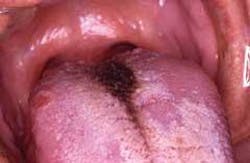
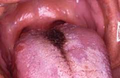
A 42-year-old male visited a dental office for a routine check-up and prophylaxis. Oral examination revealed a brownish discoloration of the dorsal tongue.
History
The patient first noticed the discoloration of the tongue approximately three months earlier. The patient denied any pain or discomfort associated with the lesion. When questioned about a history of smoking, the patient reported smoking one pack of cigarettes per day for the past 10 to 15 years.
At the time of the dental appointment, the patient's medical history was reviewed and included no significant positive findings. The patient was not taking medications of any kind at the time of the examination. The patient's past dental history included sporadic dental examinations and routine dental treatment. The patient's last dental appointment was approximately two years earlier for a dental prophylaxis.
Examinations
The patient's vital signs were obtained and all found to be within normal limits. Physical examination of the head and neck regions revealed no enlarged or palpable lymph nodes. No significant extraoral findings were noted.
Intraoral examination revealed a localized brown, matted appearance at the midline of the posterior dorsal tongue (see photo). The area exhibited a hairy appearance and could not be removed by wiping or scraping.
Clinical diagnosis
Based on the clinical information presented, which one of the following is the most likely clinical diagnosis?
◊ hairy tongue
◊ hairy leukoplakia
◊ central papillary atrophy
◊ median rhomboid glossitis
◊ hyperplastic candidiasis
Diagnosis
• hairy tongue
Discussion
As the name "hairy tongue" suggests, this is an abnormal condition in which the dorsal tongue has a hairy appearance. The hairy appearance results from the elongation and hyperkeratosis of the filiform papillae.
The exact cause of the elongation of the filiform papillae is unclear, although a number of precipitating factors have been suggested.
Such precipitating factors include the use of antibiotics, the use of oxidizing mouthwashes or antacids, general debilitation, poor oral hygiene, heavy smoking, and radiation therapy.
Clinical features
Hairy tongue occurs most frequently in males over the age of 30. The lesion typically appears as a matted plaque with a hairy appearance. This lesion is only found on the dorsal tongue; the lesion usually begins on the posterior tongue near the foramen cecum and then spreads laterally and anteriorly.
Hairy tongue may appear white, yellow, brown or black, depending upon the involved extrinsic factors (tobacco or food, for example) and intrinsic factors (chromogenic organisms, for example).
Diagnosis
The appearance of hairy tongue is characteristic. No biopsy is required. This condition is sometimes confused with hairy leukoplakia because of its similar name. It is important to note that hairy tongue and hairy leukoplakia are two distinct and separate lesions. Hairy leukoplakia is a white plaque found on the lateral borders of the tongue that is caused by the Epstein-Barr virus and is seen in association with immunosuppression.
Treatment
Hairy tongue is a benign and self-limiting condition. Empirical treatment recommendations include brushing or scraping the tongue, improving oral hygiene, and discontinuing any potential precipitating factors. In most instances, the tongue returns to its normal state subsequent to physical debridement and proper oral hygiene.
Joen Iannucci Haring, DDS, MS, is a professor of clinical dentistry, Section of Primary Care, The Ohio State University College of Dentistry.

