by Joen Iannucci Haring
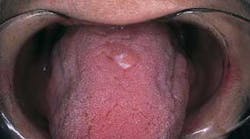
A 58-year-old female visited a dental office for fabrication of a new denture. During the oral exam, a small lump was noted on the dorsal tongue.
History
The patient stated that she had been aware of the lump for several years and described the lump as painless. The patient claimed that the lesion did not appear to increase in size and denied any injury or irritation to the involved area.
At the time of the dental appointment, the patient appeared to be in an overall good state of health. The patient's medical history was reviewed and no significant health problems were noted.
Examinations
The patient's vital signs were all found to be within normal limits. Examination of the head and neck region revealed no enlarged or palpable lymph nodes. No significant or unusual findings were discovered during the extraoral examination.
Intraoral examination revealed a sessile mass on the dorsal surface of the tongue (see photo). The lesion measured approximately 1.0 centimeters in diameter. The surface of the lesion appeared intact and white-pink in color. Palpation revealed a firm, non-movable, well-circumscribed mass. The lesion was described as painless when compressed. Further oral examination revealed no other lesions present.
Clinical diagnosis
Based on the clinical information available, which of the following is the most likely diagnosis?
o granular cell tumor
o irritation fibroma
o lipoma
o neurofibroma
o neurilemoma
Diagnosis
• granular cell tumor
Discussion
The granular cell tumor is an uncommon, benign soft-tissue tumor. As the name suggests, the cells of this tumor exhibit a granular appearance when examined histologically. The cause of the granular cell tumor is uncertain; however, evidence suggests that it is neural in origin. The granular cell tumor may occur in the oral cavity or on the skin.
Clinical features
The granular cell tumor most often occurs between the ages of 30 and 60. There is a 2:1 female predilection. The granular cell tumor usually occurs as a nodule, or sessile submucosal swelling. When palpated, the granular cell tumor feels firm, non-movable, and well-circumscribed. When compressed, the granular cell tumor is painless.
The surface of the granular cell tumor is typically intact and may appear either pink, yellow, or white in color. Although the granular cell tumor may appear in any intraoral location, it is most often found on the dorsal tongue. The granular cell tumor is a slow-growing neoplasm and seldom exceeds two centimeters in diameter. Occasionally, this lesion may occur in multiples.
Diagnosis
Based on the clinical features, a granular cell tumor may be confused with other lesions such as the irritation fibroma, neurofibroma, neurilemoma, or lipoma. A biopsy and histologic examination of such lesions establishes a definitive diagnosis. When examined histologically, the granular cell tumor exhibits large, oval cells with granular cytoplasms.
Treatment
The granular cell tumor does not disappear spontaneously or regress over time. The treatment of choice for the granular cell tumor is surgical excision. Recurrence of this lesion is not expected.
Joen Iannucci Haring, DDS, MS, is a professor of clinical dentistry, Section of Primary Care, The Ohio State University College of Dentistry.


