The Uvula
By Nancy W. Burkhart, RDH, EdD
Your patient today is 33-year-old Maggie who just had some restorative work completed by the doctor at your office during the past week. Maggie is in for her regular maintenance appointment. She was last seen by you during an appointment one year ago.
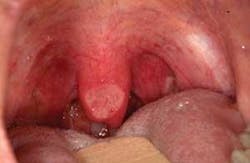
As you begin to talk with Maggie, she tells you that she was viewing her new restorative work in a mirror and believes that some problem has occurred in her posterior throat region. She reports that the area has been irritated since the procedure was completed last week. Maggie asks you if her tonsils and “whatever that thing is that hangs down in my throat” appears irritated. Maggie is referring to the uvula (see Figure 1).
She then asks you the following questions.
- What is the uvula?
- What function does the uvula provide?
- Does my uvula appear irritated to you?
Diagnosis: Maggie has a normal uvula that has an aphthous ulcer present. This is an unusual location but probably has no connection to her previous restorative work and is not due to trauma of the uvula. Because of the location on the uvula, patients may not even know that such a lesion is present other than the discomfort that may be noticed in that area.
Etiology: The uvula is a pendulum type process that extends from the midline of the soft palate and is strangely enough — a unique structure to humans. Although some animals have a similar structure that is less developed, the uvula in humans is very unique in both structure and form. The uvula is known to secrete both mucous and serous fluids that provide lubrication.
Pathogenesis: It has been suggested that the uvula is instrumental in draining the mucous secretions flowing from the nasal cavity, directing the secretions to the back of the tongue, and lubricating the base of the tongue. Other functions include protection for the eustachian tubes, immunological functions, protection of the throat from hot foods and liquids, and some authors have reported no known functions.
In early years, the uvula was removed with the tonsils because of the possibility of promoting infections. The uvula is removed in certain cultures because it is believed that uvulectomy cures disease, encourages better breast feeding, as well as an entire realm of “curative” type beliefs (Mani, 1984). This may include “malformed” uvulas that are often removed as well.
Modern science has since documented the function of the uvula as being beneficial; however, the uvulectomy is still ritually performed in some cultures on infants and the young.
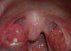
Perioral and intraoral characteristics: The uvula may have several different shapes and may vary in appearance with some individuals. The uvula presented in Figure 2 appears more rounded than the usual elongated uvula.
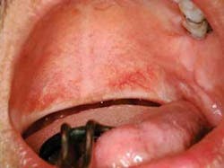
The uvula in Figure 3 is termed a “bifid uvula” and is found in approximately one in every 80 Caucasian individuals with less frequency seen in African Americans (Neville et al.). Both Asian and American Indian populations have a much higher rate of 1 in 10. Bifid uvula is the most minimal form of the cleft palate and results because of the failure of fusion in the maxillary and median nasal processes.
The uvula presented in Figure 4 appears to be the commonly seen uvula, but the bulbous tonsils seen in the slide are filled with food debris, bacteria, and concretions. The commonly used term for this condition related to the tonsils is “cryptic tonsils,” and patients often have the tonsils removed because of the ongoing problems related to this condition. The concretions become hardened and also promote halitosis in the individual.
Distinguishing characteristics: The uvula has been mentioned throughout history, from the writings of Hippocrates and Aristotle in the 4th century BC to the present scientific publications.
This structure is believed to have multiple functions. It is believed that the uvula plays a crucial role in sounds and speech. Languages such as Arabic, French, German, and Hebrew use consonants that are affected by the function of the uvula. Some West African languages and tribes who use a “clicking” sound to communicate involve the uvula in the productions of sounds (Back et al.). For the most common languages such as English, no effect is suffered when the uvula is removed. The uvula is also involved in the flow of fluid and food directing these to one side of the pharyngeal areas — thereby prohibiting entrance into the nasal passages.
Significant microscopic characteristics: The uvula is composed of connective tissue, glandular tissue, and diffused, interdigitated muscle fibers. Interestingly, the uvula has several types of thin saliva that are secreted comprising both serous and mucous components. The secretion is mostly fluid and lubricates the postpharyngeal areas, released when needed by muscle contraction (Finklestein et al., 1992).
Most of the soft palate has mucous glands that produce a thicker type of secretion. It has been demonstrated that the uvula has the ability to move backward and forward during certain functions releasing the fluid when needed and basting the pharynx with lubrication (Back et al.). This lubrication is known to aid in speech by lubricating the vocal cords.
Dental implications: The uvula may sometimes become enlarged due to several factors. Infections related to the tonsils, bacterial/viral infections in the throat, allergic type responses, and trauma-related causes. Figure 1 is a good example of the aphthous lesion that has occurred on the uvula presented in this case. The uvula may also be completely removed (uvulectomy) for reasons related to snoring and sleep apnea as seen in Figure 5. Vibration may occur during sleep, producing a snoring sound.
Other techniques alter the uvula as well, such as radio-frequency ablation (RFA) that causes the uvula to heal with scarring and then become less mobile. Because of the lubricating qualities of the uvula, many patients who have had a uvulopalatoplasty (LAUP), may report complaints of dryness in the posterior/throat areas. Recommending products and procedures to lessen xerostomia assists the patient in reducing this discomfort.
Treatment: Uvulas come in many shapes and sizes. When a patient asks why they even have one, perhaps some patient education related to the structure will be appreciated. The posterior pharyngeal region is often a forgotten one in our oral exam, and it is an important part of the total oral cancer screening. Oral cancer does occur on the uvula and the tonsils, and early detection is especially crucial in this region.
These areas are sometimes left for the ear, nose, and throat specialist (otolaryngologist) — overlooked by the dental community. Every patient should be checked at each visit during the oral cancer exam, and referred when unusual findings need further evaluations. Your patients will appreciate it!
About the Author
Nancy Burkhart, RDH, EdD, is an adjunct associate professor in the Department of Periodontics at Baylor College of Dentistry and Texas A & M Health Science Center in Dallas. Nancy is also a cohost of the International Oral Lichen Planus Support Group through Baylor (www.bcd.tamhsc.edu/lichen). She is the coauthor of General and Oral Pathology for the Dental Hygienist, published by Lippincott Williams & Wilkins in Baltimore, which was released in October 2007. She can be contacted at [email protected].
References:
- Back GW, Nadig S, Uppal S, Coatesworth AP. Why do we have a uvula?: literature review and a new theory. 2004, Clin Orolaryngol. 29, 689-693.
- Finkelstein Y, Meshorer A, Talmi Y, Zohar Y, Brenner J, Gal R. The riddle of the uvula. Otolaryngol Head Neck Surg 1992;107:444-450.
- Manni JJ. Uvulectomy, a traditional surgical procedure in Tanzania. Ann. Trop Med. Path. 1984; 78, 49-53.
- Neville BW, Damm DD, Allen CM, Bouquot JE. Oral & Maxillofacial Pathology. WB Saunders Co. Philadelphia, 1995.
- Delong L, Burkhart NW. General and Oral Pathology for the Dental Hygienist. Lippincott, Williams & Wilkins. Baltimore, 2007.


