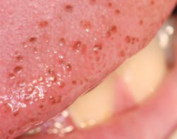
Pigmented fungiform papillae, or papillary tip melanosis
Pigmented fungiform papillae are sometimes located on the tip, lateral border, or dorsum surface of the tongue and intertwined with the filiform papillae. The fungiform papillae are involved in taste and can be very prominent in some individuals. They usually appear as a darker pink color (figure 1). Although black or brown pigmentation occurs in some patients, it is not a frequent occurrence.
When found in people with darker pigmented skin, pigmented fungiform would be considered a variant of normal oral pigmentation. In many cases, patients can remember the pigmentation as being present from childhood, and this pigmentation is usually evident during the first two decades of life. Black individuals seem to be more susceptible to darker pigmented fungiform papillae, but this is separate from racial pigmentation since some black individuals have racial pigmentation in other areas of the mouth but not on the tongue or tongue tip.
Pigmented areas in the mouth are not uncommon, but there is always a concern regarding oral pigmentation because of the possibility of melanoma or other serious pathology that should not be missed. This is often a dilemma for the dental clinician because sometimes determining the etiology is not that easy. Additionally, physicians do not usually include a total oral examination as part of patients’ physical examinations. Lambertini et al. point out that the dermatology literature is often somewhat void of oral disease signs, and oral exams are not included routinely in dermatological exams.1 Certain systemic disease states are associated with oral pigmentation, such as Peutz-Jeghers syndrome, Laugier-Hunziker syndrome, McCune-Albright syndrome, Addison’s disease, neurofibromatosis, and others. Pigmentation and melanosis (melanotic macules) may occur for a variety of reasons and may appear in multiple or single locations in the mouth and also on the lips. Melanocyte proliferation or increases in melanin production may cause changes in the oral tissue appearance.
In another article, Lambertini et al. discuss if and when clinicians should consider biopsy.2 The authors classify oral pigmentation into two major groups:
- Focal—e.g., amalgam tattoo, melanocytic nevi, melanoacanthoma, and melanosis
- Diffuse—e.g., racial/physiological pigmentation, smoker’s melanosis, drug-induced hyperpigmentation, postinflammatory hyperpigmentation, and pigmentation associated with disease states
The authors do point out that some pigmentations may not be easily diagnosed or fit the usual patterns.2 In the case of oral melanoma, early and prompt diagnosis is crucial to recovery. Oral melanoma has a worse prognosis than the cutaneous forms. A careful review of medications and health histories is needed in reaching an etiology of the pigmentations that are noted. The authors encourage colleagues to conduct a thorough examination of the oral cavity during dermatologic exams.2
Some patients present with what is termed “papillary tip melanosis,” and this is simply small brown, red, or dark pink focal pigmented specks on the tip or lateral border of the tongue. These pigmented areas are on the fungiform papillae. They usually are located on the top portion of the fungiform papillae.
The authors of a paper titled “Papillary tip melanosis (pigmented fungiform lingual papillae)” discuss a patient at their clinic who exhibits these fungiform pigmentations.3 Adibi et al. suggest the term PTM for papillary tip melanosis is more accurate and less confusing.3 The authors explain that the “bumps,” even though they are pigmented, can probably be recognized in childhood or at birth, and no treatment is needed. Pigmented fungiform papillae often go unnoticed, but any discoloration should be identified and documented by clinicians. Pigmented lesions should also be examined during future visits for any relevant changes. The tip of the tongue is not a common site for pigmentation, but the location is highly visible to the clinician during an exam. Although racial pigmentation does occur with some patients on the tip of the tongue, the authors state that the fungiform papillae are not typically involved (figures 2 and 3).3Oral melanosis, in general, may be produced by medications as well. Known medications associated with oral melanosis include tranquilizers, antibiotics, antifungals, antimalarials, arthritis medications, and hormone therapy medications. Of course, tobacco use is associated with smoker’s melanosis.
When pigmented areas are questionable, and melanoma or other serious physiological diseases may be a differential diagnosis, biopsy and further tests may be needed. Dermatoscopy is used to confirm papillary tip melanosis. A rose petal–like appearance with a very distinct pattern can be seen in the fungiform papillae viewed microscopically. This technique is used to confirm papillary tip melanosis, along with other tests when needed.4–6 Papillary tip melanosis is a benign entity, but sometimes confusion exists clinically.
As always, continue to ask good questions, and listen to your patients!
References
1. Lambertini M, Patrizi A, Ravaioli GM, Dika E. Oral pigmentation in physiologic conditions, post-inflammatory affections and systemic diseases. G Ital Dermatol Venereol. 2018;153(5):666-671. doi:10.23736/S0392-0488.17.05619-X.
2. Lambertini M, Patrizi A, Fanti PA, et al. Oral melanoma and other pigmentations: when to biopsy? J Eur Acad Dermatol Venereol. 2018;32(2):209-214. doi:10.1111/jdv.14574.
3. Adibi S, Suarez P, Bouquot JE. Papillary tip melanosis (pigmented fungiform lingual papillae). Tex Dent J. 2011;128(6):572-573,576-578.
4. Ghigliotti G, Chinazzo C, Parodi A, Rongioletti F. Pigmented fungiform papillae of the tongue: The first case in an Italian woman. Clin Exp Dermatol. 2017;42(2):206-208. doi:10.1111/ced.13006.
5. Docx MKF, Vandenberghe P, Govaert P. Pigmented fungiform papillae of the tongue (PFPT). Int J Clin Lab Med. 2016;71(2):117-118. doi:10.1179/2295333715Y.0000000067.
6. Pinos-León V. Dermoscopic features of pigmented fungiform papillae of the tongue. Actas Dermosifiliogr. 2015;106(7):527-602. doi:10.1016/j.adengl.2015.06.018.
NANCY W. BURKHART, EdD, BSDH, AFAAOM, is an adjunct associate professor in the Department of Periodontics-Stomatology at Texas A&M University College of Dentistry. Dr. Burkhart is founder and cohost of the International Oral Lichen Planus Support Group (dentistry.tamhsc.edu/olp/) and coauthor of General and Oral Pathology for the Dental Hygienist, in its third edition. She was awarded an affiliate fellow status in the American Academy of Oral Medicine in 2016. She was awarded the Dental Professional of the Year award in 2017 through the International Pemphigus and Pemphigoid Foundation and is a 2017 Sunstar/RDH Award of Distinction recipient. She can be contacted at [email protected].
About the Author
Nancy W. Burkhart, EdD, MEd, BSDH, AAFAAOM
NANCY W. BURKHART, EdD, MEd, BSDH, AAFAAOM, is an adjunct professor in the Department of Periodontics-Stomatology, College of Dentistry, Texas A&M University, Dallas, Texas. She is founder and cohost of the International Oral Lichen Planus Support Group (dentistry.tamhsc.edu/olp/) and coauthor of General and Oral Pathology for the Dental Hygienist, now in its third edition. Dr. Burkhart is an academic affiliate fellow with the American Academy of Oral Medicine, where she also serves as chair of the Affiliate Fellowship Program Committee. She was awarded the Dental Professional of the Year in 2017 through the International Pemphigus and Pemphigoid Foundation, and she is a 2017 Sunstar/RDH Award of Distinction recipient. Her professional interests are in the areas of oral medicine and the relationship between oral and whole-body health, with a focus on mucosal disease and early oral cancer detection. Contact her at [email protected].


