Muddy flooding and sinkholes: Understanding the difference between caries and erosive tooth wear
Does a sinkhole equate to tooth destruction? What does muddy flooding have to do with oral health? Conceptually, these two environmental phenomena are perfect parallels to the damage that can occur in the oral cavity.
Caries and erosion
Dental professionals are expected to recognize damage to hard tooth structure. It is easy to lump all tooth destruction under one large umbrella—caries—but in reality, there are two distinct types of loss: caries and erosive tooth wear.1
Caries is not the same as erosive tooth wear. Caries is a pH-mediated disease and an infection initiated by a variety of microbes.2-4 In contrast, erosion is an oral condition that does not involve microbes. Researchers now describe erosion as erosive tooth wear, a term that more accurately describes the complexity of the condition.⁵ The etiology of caries and erosion are simply different. So are the prevention and intervention strategies for each condition.
Caries—the microbial culprits
Caries microbes are housed in plaque biofilm, the complex microbial community attached to tooth surfaces. The slimy portion of the biofilm, which is a mucopolysaccharide, provides protection for this dynamic microbial community.6-9 Nutrients and waste products flow throughout the community via the structural dynamics created by the microbes.
Caries microbes are classified as acidogenic or aciduric. Acidogenic microbes are infection front-runners. These initial colonizers metabolize fermentable carbohydrates. Acidogenic microbes produce copious amounts of acid, creating an environmental condition that supports their proliferation.10
The caries process is further complicated by a different group of bacteria known as aciduric microbes. These microbes do not set off the initial acid attack, but when confronted with sustained low pH levels, aciduric microbes adapt their genetic makeup to survive an acidic environment. After surviving the pH drop, aciduric microbes become acid producers as well.10
Originally, scientists thought caries microbes were limited to a few bacteria, but over the last decade, the conversation has become deeper. Science is now demonstrating that the caries infection is much more complex than previously thought.11 Aggressive caries infections may also have a fungal component. Candida albicans, a common oral microbe, is a smoking gun in the caries infection. Candida grows very quickly and is a heavy acid producer, thus contributing the continued low pH levels necessary for caries.12-15
Further complicating the situation—different tooth surfaces
For years, scientists thought that Streptococcus mutans and Lactobacillus were the chief troublemakers. While these microbes are found in most carious lesions, research now indicates that caries is a highly complex infection and different tooth surfaces support different mixes of microbes—two key concepts in our scientific understanding of this disease.16-18
Caries is an all-inclusive term, indicating destructive tooth lesions that have a microbial component. Stop to consider that all caries infections are not the same. Consider a cavitated occlusal carious lesion versus a dark, leathery carious lesion on an exposed root surface. To further complicate the discussion, think about the appearance of a white spot lesion on the facial surface of a tooth. None of these three lesions has the same presentation; instead, they each indicate the presence of a different caries etiology.16,17
Current research tells us that the microbial mix in different carious lesions differs. White spot lesions are early carious lesions in the enamel that has not collapsed inward. S. mutans predominates in enamel caries. While Lactobacillus is the chief player in root caries, proteolytic microbes are also present. Collagen, a major component of dentin, is a major source of protein in the human body, and proteolytic microbes damage protein.16,17
Erosion—a chemistry lesson
Erosion is defined as a progressive loss of tooth structure not involving microbes. Acid erosion occurs when the outermost layers of the tooth surface are softened via direct contact with substances that have a low pH.19 Frequent and prolonged exposure over time eventually results in the breakdown of the tooth surface,5 creating a phenomenon called melting tooth syndrome.
There are additional sources of acid. One’s diet can play a direct role in the development of erosive tooth wear. The most obvious extrinsic culprits are acidic beverages and foods,20-23 but medications and supplements can also have low pH levels.24-27 Additional acid exposures include regurgitation in any form. A short list includes gastritis and gastroesophageal reflux disease (GERD), eating disorders such as bulimia, chemotherapy, pregnancy, and medication side effects. Other conditions can play a role as well.
In addition to low pH values, titratable acidity (TA) plays a role in the impact of any acidic product. pH values are a snapshot at a particular moment in time. The pH refers to the actual acidity of a product, but pH alone does not tell the whole story. TA refers to the effort it takes to return to a neutral environment.
TA is a numerical value—not a measurement in time. For example, the pH value of a legacy cola drink is 2.5, while the pH of a popular energy drink is around 3.6. If the pH were the only known value, then one could conclude that the cola drink has a much higher erosion potential. However, that is simply not the case. Energy drinks contain multiple organic acids. The TA of the cola is 18.3 and 51.9 for the energy drink. It takes nearly three times more effort to neutralize the energy drink. While the pH of the energy drink is higher, its erosive effect is much more profound due to the higher TA value.23
Erosion is further exacerbated by intake frequency, amount of time an acidic product remains in contact with the tooth structure, and the body’s ability to neutralize the acid.
A more complex time bomb
Fermentable carbohydrates further compound the acidic impact. In this case, the microbes are being fed and the acidic metabolic waste products are increasing damage to the tooth. While foods and beverages are obvious carbohydrate sources, many medications and supplements are also rich sources of carbohydrates.28-31 The product platform also plays a role. Thick, sticky liquids and gummy gel formats keep medications and supplements in contact with tooth structure for protracted periods of time.
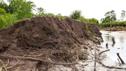
Creating a visual story
Most people are visual learners. Visual storytelling is a great way to convey complex concepts. This is where muddy flooding and sinkholes come into play. It’s easy to find web-based examples of both. Both phenomena are manifestations of physical loss, which can be great analogies when trying to explain the difference between erosion and caries.
Agronomists and soil experts know all about muddy flooding. So do those who live along riverbanks or lakeshores. Muddy flooding occurs when water either rises above the banks or shorelines,32 or when there is excessive rainfall. During muddy flooding, the ground gets soft and soil particles erode from the surface and become suspended in the water. The soil particulate is now eroded from the original surface. All of the action takes place on the surface of the land, just as the enamel and dentin structure erode over time due to surface softening from acid attacks.
Carious lesions are like sinkholes. Biofilm microbes produce lactic acid, which percolates through the enamel surface and subsequently weakens the dentin substructure. A white spot lesion is an early indication that subsurface damage is taking place. As long as the enamel surface remains intact, it is possible to reverse the process and remineralize the underlying tooth structure. Once cavitation has occurred, a carious lesion will need more definitive treatment.
Final thoughts
Oral conditions that damage and destroy tooth structure can’t just be classified as caries. Caries is not the only problem. Erosion plays an increasingly important role. The destruction is complex. The loss of tooth structure can be stopped if identified early enough and reversed if there is still sufficient structural integrity.
Information and education are the key. Print out photos of muddy flooding and sinkholes. Laminate them and use the visual images to create a conversation with your patients. Initiate meaningful conversations with patients and be willing to put the old proverb—out of sight, out of mind—to rest by utilizing new visual concepts and storytelling.
References
- Marsh PD. Are dental diseases examples of ecological catastrophes? Microbiology. 2003;149(2):279-294. doi:10.1099/mic.0.26082-0
- Marsh PD. Microbiology of dental plaque biofilms and their role in oral health and caries. Dent Clin North Am. 2010;54(3):441-454. doi:10.1016/j.cden.2010.03.002
- Takahashi N, Nyvad B. Caries ecology revisited: microbial dynamics and the caries process. Caries Res. 2008;42(6):409-418. doi:10.1159/000159604
- Cavalcanti YW, Bertolini MM, da Silva WJ, Del-Bel-Cury AA, Tenuta LMA, Cury JA. A three-species biofilm model for the evaluation of enamel and dentin demineralization. Biofouling. 2014;30(5):579-588. doi:10.1080/08927014.2014.905547
- Lussi A, Schlueter N, Rakhmatullina E, Ganss C. Dental erosion--an overview with emphasis on chemical and histopathological aspects. Caries Res. 2011;45 Suppl 1:2-12. doi:10.1159/000325915
- Cugini C, Shanmugam M, Landge N, Ramasubbu N. The role of exopolysaccharides in oral biofilms. J Dent Res. 2019;98(7):739-745. doi:10.1177/0022034519845001
- Koo H, Falsetta ML, Klein MI. The exopolysaccharide matrix: a virulence determinant of cariogenic biofilm. J Dent Res. 2013;92(12):1065-1073. doi: 10.1177/0022034513504218
- Klein MI, Hwang G, Santos PHS, Campanella OH, Koo H. Streptococcus mutans-derived extracellular matrix in cariogenic oral biofilms. Front Cell Infect Microbiol. 2015;5:10. doi:10.3389/fcimb.2015.00010
- Cavalcanti YW, Bertolini MM, da Silva WJ, Del-Bel-Cury AA, Tenuta LMA, Cury JA. A three-species biofilm model for the evaluation of enamel and dentin demineralization. Biofouling. 2014;30(5)579-588. doi:10.1080/08927014.2014.905547
- Burne RA, Zeng L, Ahn SJ, et al. Progress dissecting the oral microbiome in caries and health. Adv Dent Res. 2012;24(2):77-80. doi:10.1177/0022034512449462
- Johansson I, Witkowska E, Kaveh B, Holgerson PL, Tanner ACR. The microbiome in populations with a low and high prevalence of caries. J Dent Res. 2016;95(1):80-86. doi:10.1177/0022034515609554
- Jenkinson HF, Douglas LJ. Interactions between Candida species and bacteria in mixed infections. In: Brogden KA, Guthmiller JM, eds. Polymicrobial Diseases. ASM Press; 2002:chap 18.
- Falsetta ML, Klein MI, Colonne PM, et al. Symbiotic relationship between Streptococcus mutans and Candida albicans synergizes virulence of plaque biofilms in vivo. Infect Immun. 2014;82(5):1968-1981. doi:10.1128/IAI.00087-14
- Hwang G, Liu Y, Kim D, Li Y, Krysan DJ, Koo H. Candida albicans mannans mediate Streptococcus mutans exoenzyme GtfB binding to modulate cross-kingdom biofilm development in vivo. PLoS Pathog. 2017;13(6):e1006407. doi:10.1371/journal.ppat.1006407
- Khoury ZH, Vila T, Puthran TR, et al. The role of Candida albicans secreted polysaccharides in augmenting Streptococcus mutans adherence and mixed biofilm formation: in vitro and in vivo studies. Front Microbiol. 2020;11:307. doi:10.3389/fmicb.2020.00307
- Aas JA, Griffen AL, Dardis SR, et al. Bacteria of dental caries in primary and permanent teeth in children and young adults. J Clin Microbiol. 2008;46(4):1407-1417. doi:10.1128/JCM.01410-07
- Preza D, Olsen I, Aas JA, Willumsen T, Grinde B, Paster BJ. Profiles of root caries in elderly patients. J Clin Microbiol. 2008;46(6):2015-2021. doi:10.1128/JCM.02411-07
- Kuramitsu HK, Wang BY. The whole is greater than the sum of its parts: dental plaque bacterial interactions can affect the virulence properties of cariogenic Streptococcus mutans. Am J Dent. 2011;24(3):153-154.
- Schlueter N, Luka B. Erosive tooth wear - a review on global prevalence and on its prevalence in risk groups. Br Dent J. 2018;224(5):364-370. doi:10.1038/sj.bdj.2018.167
- Carvalho TS, Schmid TM, Baumann T, Lussi A. Erosive effect of different dietary substances on deciduous and permanent teeth. Clin Oral Investig. 2017;21(5):1519-1526. doi:10.1007/s00784-016-1915-z
- Barbour ME, Lussi A. Erosion in relation to nutrition and the environment. Monogr Oral Sci. 2014;25:143-154. doi:10.1159/000359941
- West NX, Hughes JA, Addy M. The effect of pH on the erosion of dentine and enamel by dietary acids in vitro. J Oral Rehabil. 2001;28(9):860-864.
- Owens BM, Kitchens M. The erosive potential of soft drinks on enamel surface substrate: an in vitro scanning electron microscopy investigation. J Contemp Dent Pract. 2007;8(7):11-20.
- Hellwig E, Lussi A. Oral hygiene products, medications and drugs - hidden aetiological factors for dental erosion. Monogr Oral Sci. 2014;25:155-162. doi:10.1159/000359942
- Delgado AJ, Olafsson VG, Donovan TE. pH and erosive potential of commonly used oral moisturizers. J Prosthodont. 2016;25(1):39-43. doi:10.1111/jopr.12324
- Delgado AJ, Olafsson VG. Acidic oral moisturizers with pH below 6.7 may be harmful to teeth depending on formulation: a short report. Clin Cosmet Investig Dent. 2017;9:81‐83. doi:10.2147/CCIDE.S140254
- Tayee P, Hsu A, Messer R, et al. Evaluation of pH values of products managing xerostomia. Dept. Oral Biology, Augusta University, Augusta, GA. 2015. Accessed July 8, 2020. https://cdn.shopify.com/s/files/1/0035/3832/files/Tayee_et_al-_Ph_values_of_xerostomia_products_2015.pdf
- Lussi A, Carvalho TS. Analyses of the erosive effect of dietary substances and medications on deciduous teeth. PLoS One. 2015;10(12):e0143957. doi:10.1371/journal.pone.0143957
- Scadding G. Pediatric allergy medications: review of currently available formulations. Curr Med Res Opin. 2009;25(8):2069-1079. doi:10.1185/03007990903116875
- Al Humaid J. Sweetener content and cariogenic potential of pediatric oral medications: a literature. Int J Health Sci (Qassim). 2018;12(3):75-82.
- Babu KLG, Doddamani GM, Naik LRK, Jagadeesh KN. Pediatric liquid medicaments - are they cariogenic? An in vitro study. J Int Soc Prev Community Dent. 2014;4(2):108-112. doi:10.4103/2231-0762.137637
- Boardman J, Vandaele K. Soil erosion, muddy floods and the need for institutional memory. Area. 2010;42(4):502-513. Accessed April 8, 2020. www.jstor.org/stable/40890908
Anne Nugent Guignon, MPH, RDH, CSP, has received numerous accolades over four decades for mentoring, research, and guiding her profession. As an international speaker and prolific author, Guignon focuses on the oral microbiome, erosion, hypersensitivity, salivary dysfunction, ergonomics, and employee law issues. She may be contacted at [email protected].
About the Author

Anne Nugent Guignon, MPH, RDH, CSP
ANNE NUGENT GUIGNON, MPH, RDH, CSP, has received numerous accolades over four decades for mentoring, research, and guiding her profession. As an international speaker and prolific author, Guignon focuses is on the oral microbiome, erosion, hypersensitivity, salivary dysfunction, ergonomics, and employee law issues. She may be contacted at [email protected].