Embracing Digital Radiography
QUALITY DIGITAL IMAGES ARE AVAILABLE, AND THEN THE SKY IS THE LIMIT FOR DIAGNOSTIC USE
BY Ann-Marie DePalma, RDH, MEd, FADIA, FAADH
The past several years have seen an increase in the number of dental practices that have adopted digital radiography.
The cost versus benefit ratio and the return on investment for practices have greatly increased with patients expecting the latest technology from their dental teams. The initial investment the practice must make in dental hardware, software, and imaging devices is often a deterrent, but in the long run with the elimination of film and solution purchases and solution disposal fees, the practice can realize savings. Additionally, when digital imaging is incorporated into the patient visit and the patient understands the need for treatment with better visualization of the disease process, case acceptance can increase.
Even so, many dental team members are hesitant to fully embrace the digital radiography revolution. Many hygienists feel knowledgeable and comfortable with using film, and the idea of change can spark fear in the hearts of many seasoned clinicians. Nevertheless, with a few simple tweaks of traditional radiographic skills, hygienists can take radiographs with digital tools that offer exceptional diagnostic quality. This article will review basic radiographic principles to assist the clinician in obtaining quality digital images.
Digital radiography has numerous advantages over film-based imaging in that the clinician has the ability to view images on the screen, archive images within the practice-management software for future viewing, enhance the images for better diagnostic ability, and potentially reduce the radiation exposure for the patient.
A diagnostic quality image is the goal of both film and digital imaging, and both rely on placement and exposure techniques and patient management skills. Diagnostic quality images involve good contrast, density, sharpness, and resolution with low noise and distortion. Diagnostic details also involve images that contain several shades of gray, sharp margins, and with no image distortion.
Factors that also influence the image quality include:
• The X-ray generator and settings
• The use of holders/barriers
• The sensor and cone placement
Following the ALARA principle (As Low As Reasonably Achievable) and the ADA Guidelines for Dental Radiographic Examinations, Recommendations for Patient Selection and Limiting Radiation Exposure, clinicians are expected to use the guidelines in conjunction with professional judgment as to when to expose a patient to radiographs. A review of the patient's medical and dental history with a risk-and-need assessment is essential for determining a patient's need for digital imaging.
Sensors
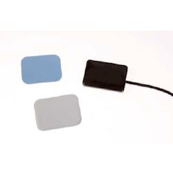
There are two basic types of digital imaging receptors: photostimulable phosphor plates (PSP) and wired or wireless charge coupled devices (CCD) or complementary metal-oxide semiconductor device (CMOS). The CCD and CMOS devices convert light to electrons using different types of technology to create pixel images that are beyond the scope of this article to discuss.
Sensor sizes are comparable to film, size 0, 1, and 2. Both PSP and CCD/CMOS are reusable but can follow different infection-control routines. Plastic barriers should also be used based on the type of imaging system. It is important to follow the manufacturer's directions regarding infection-control protocols for each type of device used. Similar to film, PSPs are flexible, while CCD/CMOS are rigid, inflexible, and unforgiving in placement. PSPs must be handled carefully and scanned to view images and then erased for future use. Improper handling of PSPs can result in scratches that can cause image artifacts and result in the need for device replacement or excessive patient radiation exposure.
Rigid sensors (CCD/CMOS) are more difficult to use initially due to the learning curve of placement and exposure, but once the learning curve is achieved, they are more user-friendly. Connection issues with rigid sensor imaging occur due to the improper handling and care of the wire that connects the sensor to the imaging remote/computer. Occasionally there will be problems with the sensor itself, but that occurs less often than the wire-related issues.
Wireless sensors sometimes have issues with connections as well due to inconsistent network conductivity. Depending on the CCD/CMOS sensor used, there may also be a narrower field of imaging than film or PSP; therefore, the clinician may have to adjust placement accordingly.
PSP
CCD/CMOS
Patient acceptance
Patient acceptance and comfort is essential for successful intraoral digital imaging. Understanding the differences between film placement and sensor placement for optimum quality imaging is the first step in assuring the patient is comfortable during the radiographic process. Some sensors have rounded edges that make placement slightly more comfortable for the patient or self-adhesive foam covers can be applied over the sensor to cushion it in the mouth. Neither, however, does more to improve patient acceptance of digital imaging than educating the patient on the process. If the patient understands the process, the comfort and acceptance level are greatly improved.
Depending on the type of radiographic imaging equipment used (i.e., old X-ray units versus newer units), it is this author's opinion not to specifically state that digital is less radiation than film. I have seen old X-ray units when retrofitted for digital need to have impulse time set to close to the film exposure time pulse. The newer X-ray units, however, offer distinct decreases in radiation exposure. Lead-lined thyroid collars and aprons should also continue to be used for digital imaging, which also presents the patient with greater comfort levels. Although the National Council on Radiation Protection (NCRP) Report 145 states that lead aprons are not required, it stipulates that rectangular collimation, fast imaging receptors, patient selection criteria along with other standards must be initiated prior to exposing radiographs without lead aprons or thyroid collars. Does your office follow all of these specifications?
Similar to film imaging where obtaining the best diagnostic quality image with the least amount of radiation exposure is achieved using film-positioning devices, optimum digital imaging is achieved in the same manner. There are numerous positioning devices on the market for all types of intraoral digital images.
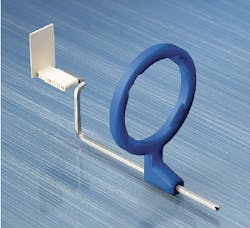
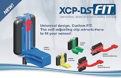
As with instrument selection and use, where there are personal and practice preference considerations for the type of instrument selected, so it is with positioning devices. Each clinician and practice must choose between a variety of positioning devices available on the market to best suit the patient and practice needs. My personal favorites may not be yours. I choose to use either disposable sticky tabs or the Rinn XCP Fit System. I find it appropriate to have both on hand since the sticky tabs are disposable and slightly less bulky than the XCP-Fits, but the Fits work better for some patients and are autoclavable.
Sticky Tabs
RinnXCP Fit
The placement and use of PSP sensors are similar to film-based principles. Whether you are using the paralleling or bisecting angle technique, you can continue to take images in a similar manner. Instead of developing film, however, the PSP sensor must be scanned into the computer software. This may involve the clinician leaving the patient similar to the time needed to develop the film, depending on where the scanner unit and computer are located.
Once the image is scanned into the system, images are available at any computer that is networked to the scanner. Depending on the software, these images may or may not be directly imported into the practice-management software or may need to be bridged to the patient's record. Once in the patient's record, they can be emailed, printed, and manipulated as appropriate. After each use, PSPs are exposed to white light to erase the image and allow reuse.
The images taken with CCD/CMOS sensors are immediately available on the computer that exposes the image as well as any other networked computer with no other intervention or time needed. Images can also be emailed, printed, and manipulated as the software allows and can be bridged if not directly imported into the patient's record.
Additionally, when using CCD/CMOS sensors, it is best to adjust the cone placement so that the cone or the ring of the placement device touches the patient's face and the wire of the sensor follows the arm of the placement device. It is this author's recommendation that readers either have an in-office team training session or take a continuing education program to understand the appropriate placement for diagnostic images based on your practice's imaging system.
Digital dentistry is here to stay. Help diagnose disease and increase case acceptance by using digital radiography to the fullest extent. You, the practice, and the patient will benefit. RDH
Tweaking sensor placement
CCD/CMOS sensors require a rethinking of technique. Since the sensors are rigid, inflexible, and bulky, using a strict paralleling or bisecting technique is difficult. I describe the use of CCD/CMOS sensors as a cross between the two techniques-not paralleling or bisecting, but halfway in between. The sensor needs to be placed more lingually or palatally toward the midline than film and may need to be more angled in order to obtain the appropriate images.
When taking premolar bitewings, the front of the sensor needs to be placed toward the opposite canine to open contacts and obtain diagnostic quality images. For patient comfort for those who have mandibular issues such as tori, or for better comfort generally, the sensor can be placed directly on the tongue.
These and other tweaks of traditional imaging techniques can provide diagnostic images and optimum patient comfort and acceptance while keeping the sensor as parallel as possible.
ANN-MARIE C. DEPALMA, RDH, MEd, FADIA, FAADH, is a Fellow of the American Academy of Dental Hygiene and the Association of Dental Implant Auxiliaries, as well as a continuous member of ADHA. She presents continuing education programs for dental team members on a variety of topics. Ann-Marie is collaborating with several authors on various books for dental hygiene and can be reached at [email protected].


