Disappearing tooth structure: What's a clinician to do about abfraction lesions?
The first time I heard the word “abfraction” was in the dental practice where I currently work, and it was about 30 years ago. My boss, Dr. Tom McDougal, mentored me in the “art and science” of identifying these interesting lesions in which the cervical portion of the tooth literally disappears. Even if you are not familiar with the word abfraction, you have looked at these in patient after patient and may have even witnessed them deepen as tooth structure disappears over time.
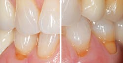
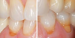
Figure 1 is an example of a typical abfraction lesion, and as is the case with many of these awkward areas of plaque retention, it is asymptomatic. Upon first glance it appears as though it should be sensitive, and certainly in some cases these are. But thanks to an incredible physiological process that happens in the face of injury, irritation, or exposure, “tertiary” or “reparative” dentin often develops to deposit additional layers of dentin to protect the pulp from hypersensitivity. While dentin is harder than bone, it is up to 10 times softer than enamel. So once exposed, it becomes vulnerable to loss of minerals from chemical exposures and abrasion, and can be plaque-retentive enough to increase caries risk.
More from the author ... Guided biofilm therapy: What is it, and why do you need it?
While the disappearing tooth structure is certainly real, the causes behind it are uncertain. More importantly, how to handle it appears to be somewhat controversial. What should registered dental hygienists advise their patients to do about their abfraction lesions? Monitor them? Restore them? Ignore them? Should they be documented? If so, how? The answers depend in part on the dentist with whom the dental hygienist works, and his or her opinion and expertise in managing abfraction lesions. Also, because these lesions are so common, a review of current literature on the subject is warranted.
Relevant data on abfractions
Enter an interesting study published in 2016 by Nascimento et al. on this topic titled, “Abfraction lesions: Etiology, diagnosis, and treatment options.”1 The Latin origin of the word abfraction means “to break away,” and refers to the theory that the occlusal compressive forces and tensile stresses create tooth flexure in the cervical area, which results in microfractures of the hydroxyapatite crystals of the enamel and dentin, and hence, a “lesion” is formed. While evidence of this process is easily seen from observing teeth that wear simultaneously in opposing arches, it is currently believed that noncarious cervical lesions (NCCLs), including abfractions, are multifactorial in their etiology.
To me, this is relevant because if there are multiple factors in the etiology, simply reducing excessive occlusal forces with something like a bite guard might focus on only one aspect of the problem. Another concern is that diagnosis and treatment planning to restore abfraction lesions depends largely on the opinion of the dentist, without a significant amount of evidence-based literature to guide his or her decisions about whether or not to restore them.
With the aging population, clinicians should expect to see an increase in NCCLs. But limited longevity of restored cervical lesions begs the question: When or should these areas of missing tooth structure be restored?
Figure 1: Abfraction lesions at the cervical portion of the tooth
Assessments first
Attempting to discover the multifactorial etiology that contributes to the disappearance of hard tooth structure at the CEJ should be a priority for clinicians. Most current literature supports the idea that mineral loss due to chemical and abrasive exposures initiates the development of a lesion, and occlusal stresses perpetuate it, literally creating a cycle of interplaying risk factors that gradually deepen the lesions over time without intervention.
Patients with occlusal wear and obvious abfraction lesions are often advised to have a customized occlusal guard fabricated to wear at night to protect against undue occlusal loads. However, the subject of flat-planed occlusal guards is a topic that is evolving in dentistry.
While they can provide beneficial protection for many patients, a recent policy statement from the American College of Prosthodontists should be a call to action to carefully consider this issue.2 This 2016 policy statement states, “Practitioners should screen patients for obstructive sleep apnea (OSA) prior to fabricating a maxillary night guard that increases the occlusal vertical dimension (OVD) without mandibular protrusion.” This reflects data that states that increasing OVD with a maxillary night guard without mandibular protrusion aggravates OSA in some patients.2 In other words, trying to protect teeth from wear by fabricating an occlusal guard might actually make sleep apnea worse in some patients, so evaluation for sleep disordered breathing, including OSA, is indicated first.
Additionally, asking patients about their dietary and beverage habits of frequent acidic exposures should help to identify those who are literally eroding away their tooth structure. They are often completely unaware of the effects of acidic foods and drinks. Clinicians should also inquire about whether or not a patient with abfraction lesions has acid reflux, while remembering that some reflux is “silent” and does not present with typical symptoms of acid indigestion, bloating, and stomach pain.
Also of importance is identifying the type of toothpaste a patient uses as it can be a clue as to why the lesions are gradually growing. The relative dentin abrasiveness (RDA) of toothpastes ranges from the low end of the spectrum, with toothpastes such as Sensodyne’s Pronamel (RDA 34) or CloSYS (RDA 53), all the way up to highly abrasive toothpastes such as Crest Pro Health (RDA 155) and Colgate Tartar Control (RDA 165). Obviously, recommending low abrasive toothpastes in the presence of abfraction lesions is indicated.
To treat or not?
When should abfractions be restored? Nascimento et al.1 provide practical recommendations in their recent publication.
- Active, cavitated, carious lesions - caries management by risk assessment (CAMBRA) is essential to determine the risks and interventions
- Cervical defects that extend subgingivally and preclude adequate plaque control
- Extensive tooth structure loss compromising the integrity of the tooth or presenting with pulpal exposure
- Persistent tooth hypersensitivity when therapeutic options have failed
- The tooth will become a prosthetic abutment
- Esthetic demands on the part of the patient
The authors remind clinicians that in the absence of strong evidence to treat, a careful risk/benefit analysis should precede restorative treatment of abfraction lesions.
This topic almost lends itself to the analogy of opening Pandora’s Box because there are other risk factors that can be identified and modified. Considerations regarding possible root coverage with grafting procedures for exposed roots and treatment of hypersensitive defects are all subjects that warrant more attention. But for the moment, here is a recap from this article:
- Clinicians need to recognize that disappearing tooth structure is multifactorial in nature.
- We owe it to our patients to identify and help modify risk factors for abfractions.
- Decisions to treat abfraction lesions should be based on evidence, a risk/benefit analysis, and the consideration of obstructive sleep apnea prior to recommending occlusal guards.
Whew! That is a lot to digest in one column. Share this with other clinicians on your team as an opportunity to open the discussion on an important topic that is not going away any time soon.
Editor's note: This content was oiginally published in 2017. It has been updated as of July 2025.
References
- Nascimento MM, Dilbone DA, Pereira PN, Duarte WR, Geraldeli S, Delgado AJ. Abfraction lesions: etiology, diagnosis, and treatment options. Clin Cosmet Investig Dent. 2016;8:79-87. doi:10.2147/CCIDE.S63465
- American College of Prosthodontists Position Statement. 2016.
About the Author
Karen Davis, BSDH, RDH
Karen Davis, BSDH, RDH, is the founder of Cutting Edge Concepts, an international continuing education company. She practices dental hygiene in Dallas, Texas. She is an independent consultant to the Philips Corporation, Periosciences, Hu-Friedy Group, and EMS. She can be reached at [email protected].


