Implant-associated diseases
What you'll learn in this article
- Why screw-retained implants lower the risk of peri-implant disease compared to cement-retained options.
- How to recognize early signs of peri-implant mucositis and prevent progression to peri-implantitis.
- Key risk factors for implant failure—and how dental hygienists can help reduce them through routine monitoring and patient education.
Listen to the article on our podcast!
Editor's note: This is part one of a two-part series.
In the last 40 years, implants have had a remarkable impact on restorative dentistry. Approximately 3 million people currently have dental implants, with an estimated 500,000 each year being placed in the United States.1-3 Dental implants have become an essential restorative option for patients with missing teeth due to a high success rate, the ability to maintain oral health, and improved ease of placement. This highlights the importance that dental hygienists be familiar with dental implants, peri-implant diseases, and implant maintenance. RDH magazine will be featuring a two-part series of articles on implants: the first will focus on peri-implant diseases, and the second will address best practices for maintaining dental implants.
To accurately identify the periodontal conditions associated with dental implants, it is necessary to understand the components of an implant, recognize the differences between screw- and cement-retained implant crowns, and understand the biological integration into the periodontium. In December 2021, the Journal of Dental Hygiene conducted a survey evaluating dental implant curriculum in dental hygiene programs. While the response sample was limited, it revealed that due to individual program constraints, there is substantial room for improvement in implant education and maintenance.4,5 This study reinforces the necessity that dental hygienists be competent, educated, confident, and up-to-date on the most current research pertaining to implants. In doing so, this renewed understanding of potential risk factors can be identified prior to the placement of dental implants and during the maintenance phase.
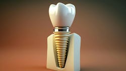
Dental implant components
Dental implants are an ideal restorative option for single or multiple missing teeth and fully edentulous patients. The implant itself mimics the root structure and allows for minimal bone resorption, preservation of the alveolar ridge, and stimulation of bone growth. Dental implant restorations can be screw-
retained, cemented, or a combination of both. There are advantages and disadvantages to a screw-retained implant versus a cement-retained implant. A screw-retained dental implant decreases the risk of developing peri-implant diseases due to residual cement. However, a screw-retained implant may not be as esthetically appealing due to a visible access point on the occlusal surface. The access point on a screw-retained dental implant allows the restorative dentist to tighten, replace, or remove the implant crown. On the contrary, a cement-retained dental implant cannot be removed without the entire implant crown being replaced. Cement-retained implants are often preferred when esthetics are a high priority for both the provider and patient.
It is important to understand the differences in the soft tissue structures of a natural tooth versus a dental implant to properly assess periodontal health. While dental implants decrease the risk of the alveolar bone becoming atrophied, the soft tissue surrounding the implant differs significantly from natural teeth. Unlike natural teeth, dental implants do not have any periodontal ligaments; therefore, the mucosal-abutment attachment can be easily harmed while probing. This can cause both bleeding and periodontal pockets deeper than 3 mm in the probed areas. If probing, less force should be applied than what is used with natural teeth.
Peri-implant diseases
Dental hygienists should understand the patient’s periodontal history, as well as assist the dentist in assessing the health of dental implants. The information collected during the patient’s appointment will provide essential information to the dentist when diagnosing peri-implant diseases. Dental hygienists should evaluate dental implants for clinical signs of inflammation and disease, such as suppuration, mobility, and bone loss.4
A recent study examined the prevalence of cement in the presence of peri-implant diseases, revealing that residual cement was present in about 80% of peri-implant diseased implants.6 Peri-implant diseases affect the supporting oral structures of the dental implant. There are two distinct peri-
implant conditions that dental hygienists need to be aware of: peri-implant mucositis and peri-implantitis.
Risk factors for peri-implant disease:
- Poor oral hygiene
- Loss of keratinized tissue
- Open contacts
- Overcontoured restorations
- Residual cement
Peri-implant mucositis
Peri-implant mucositis is an acute condition that affects the soft tissues surrounding the dental implant in approximately 40%–50% of all implants.4 Clinically, the tissue may be red, swollen, and tender. Like gingivitis, this condition is reversible, and if detected early, it is easily treated with improved home care and an increase in recall appointments. There is a direct connection between plaque control and peri-implant mucositis.7 Patients who present with bleeding upon probing, an increase in pocket depth, and red, swollen tissue, but no radiographic bone loss should be diagnosed with peri-implant mucositis. If left untreated, peri-implant mucositis is a precursor for peri-implantitis.
Peri-implantitis
Peri-implantitis is a chronic condition in which the peri-implant mucositis has progressed to peri-implantitis. This disease involves both the soft and hard tissues surrounding the dental implant and affects between 12% and 43% of implants.4 A key clinical finding with this diagnosis is radiographic bone loss, which can also be in conjunction with mobility and suppuration. Patients who smoke, have periodontal disease, poor plaque control, or systemic conditions that impact the oral cavity are at an increased risk of developing peri-implantitis and should be monitored closely.
A recent study suggests that patients with poor plaque control and limited oral accessibility had a significantly higher risk of peri-implantitis than those with cleansable implants.7 Treatment options for patients with peri-implantitis frequently require surgical intervention. Etiologic factors that may coincide with peri-implantitis should be identified as quickly as possible, including systemic health conditions. It is recommended that yearly radiographs be taken of dental implants to monitor bone levels and the apex of the implant. Close monitoring of dental implant patients will allow for a prompt diagnosis so that treatment can begin at the earliest sign of disease.
Early identification of peri-implant diseases increases the likelihood of slowing the progression of the disease process and initiating necessary treatment. It is estimated that approximately 15% of all dental implants will fail during the first 10 years.8 Educating patients on the risk factors and signs of peri-implant diseases will help reduce the risk of them developing peri-implantitis. By working together as a team with the patient and dentists, dental hygienists play in integral role in preventing peri-implant diseases and decreasing the risk of failing dental implants.
Editor's note: This article appeared in the July 2025 print edition of RDH magazine. Dental hygienists in North America are eligible for a complimentary print subscription. Sign up here.
References
- Van Witzenburg M. Implant maintenance: getting past the fear. RDH. December 1, 2019. Accessed March 1, 2025. https://www.rdhmag.com/patient-care/implant-maintenance/article/14073139/implant-maintenance-getting-past-the-fear
- Implants. American Dental Association. MouthHealthy. Accessed March 1, 2025. https://mouthhealthy.org/en/az-topics/i/implants
- What are dental implants? American Academy of Implant Dentistry. Accessed March 5, 2025. https://www.aaid-implant.org/dental-implants/what-are-dental-implants/
- Gentry AC, Gurenlian JR, Freudenthal JJ. Implant maintenance curriculum content in dental hygiene education. J Dent Hyg. 2021;95(6):36-45.
- Figuero E, Graziani F, Sanz I, Herrera D, Sanz M. Management of peri-implant mucositis and peri-implantitis. Periodontol 2000. 2014;66(1)255-273. doi:10.1111/prd.12049
- Montevecchi M, Valeriani L, Salvadori MF, Stefanini M, Zucchelli G. Excess cement and peri-implant disease: a cross-sectional clinical endoscopic study. J Periodontol. 2025:1-9. doi:10.1002/JPER.24-0510
- Scarano A, Khater AGA, Gehrke SA, et al. Current status of peri-implant diseases: a clinical review for evidence-based decision making. J Funct Biomater. 2023;14(4):210. doi:10.3390/jfb14040210
- Jemt T. Implant failures and age at the time of surgery: a retrospective study on implant treatments in 4585 edentulous jaws. Clin Implant Dent Relat Res. 2019;21(4):514-520. doi: 10.1111/cid.12753
About the Author

Melissa Van Witzenburg, MS, RDH
Melissa has been practicing dental hygiene for 23 years. She continues to pursue her passion by educating the aging population about oral health and systemic links. Melissa also works clinically in a periodontal office in the Chicagoland area. For more information, email her at [email protected].