Rethinking caries detection: Preserving enamel to prevent the restorative cycle
What you'll learn in this article
- Why traditional caries detection methods may miss early-stage disease
- The dangers of outdated probing techniques
- How emerging technologies are detecting demineralization earlier
- The difference between lagging and leading indicators in caries detection
- How caries risk assessments and personalized prevention strategies improve outcomes
In the dental hygiene program where we teach, clinical detection of caries has traditionally relied on a combination of explorer use, visual assessment, radiographs, and laser fluorescence for tooth surface analysis. As we shift toward a more preventive and minimally invasive philosophy, are these practices working and are we detecting disease early enough to save our patients’ teeth?
Incorporating remineralization strategies into our clinic protocols has opened new conversations about earlier intervention to catch demineralization before it becomes a visible white spot lesion, or worse, cavitated decay. If dental hygienists can support enamel health before irreversible damage occurs, we stand a better chance of keeping patients out of the restorative cycle altogether.
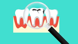
After all, this is one of the core goals of dental hygiene: to prevent disease and preserve natural tooth structure. We can visibly detect caries once a white spot lesion has developed. This means that around 200 UM of precious tooth hydroxyapatite has been lost and it’s too late.1
New ways to approach potential caries
Many of us were trained to use a sharp-ended explorer to detect “stickiness” in suspected carious lesions. However, research has shown this method can damage weakened enamel and potentially accelerate lesion progression.
In a landmark study, Ekstrand et al. found that probing with a sharp instrument can convert a noncavitated lesion (and one that’s potentially able to be remineralized) into an irreversible cavity, disrupting the surface and eliminating the chance for remineralization.2 This evidence reinforces the importance of gentle, noninvasive diagnostic approaches, particularly when combined with modern tools and preventive strategies.
For interproximal areas that cannot be manually inspected with a dental explorer, radiographs are traditionally used to screen for decay. Several research studies show that dental radiographs, such as bitewings, typically detect tooth decay only after significant demineralization has occurred. Specifically, enamel caries must penetrate approximately 230 to 410 micrometers into the enamel before becoming reliably visible on radiographs.3
In addition, the increased depth of enamel demineralization is positively correlated with rate of detection. In other words, carious lesions must progress to a more advanced stage to be reliably detected with radiographs,4 which is counterintuitive to the primary objective of preventive care.
The growth of fluoresence
Laser fluorescence from products such as DIAGNOdent is also becoming more popular, with several products now on the market using this technology to detect demineralization. When this 655 nm diode laser is directed onto a tooth's surface, its light is absorbed by metabolic byproducts of oral bacteria. These compounds then emit red fluorescence, which is captured by the device and translated into a numerical reading from 0 to 99.
Using the DIAGNOdent doesn’t have the same risks as an explorer, and laser fluorescence has a high sensitivity, meaning it’s accurate in detecting decay, but it has low specificity, meaning that it often produces false negatives. Therefore, laser fluorescence is recommended only as an adjunct device to be used alongside radiographic and clinical evaluation to definitively diagnose decay.5
Similarly, LED fluorescence stimulates metabolic products found in cariogenic bacteria, causing them to glow red, while healthy enamel glows green and shows a visual image on the screen. Other diagnostic tools include near-infrared light, which penetrates enamel deeply through transillumination. Healthy enamel will appear translucent and carious lesions stand out as dark zones.
Other popular diagnostic tools
One exciting innovation that detects demineralization with only ~30–50um of hydroxyapatite loss is BlueCheck, a paint-on diagnostic agent composed of a protein with a high affinity for hydroxyapatite. BlueCheck is a leading indicator, detecting disease earlier than can even be seen visually.
When applied to the tooth surface, BlueCheck helps highlight areas with active demineralization to provide visual cues for clinicians and patients alike. While interproximal detection can be limited, it brings early surface demineralization to light in real time, allowing it to be treated before it becomes a white spot lesion. Check this out in action on Shelley’s dental educational YouTube channel.
Two other cutting-edge disease-indicator tools are LumiCare and Calcivis. LumiCare uses a cavity-detection rinse that contains biocompatible and fluorescent starch nanoparticles that penetrate early-stage, porous enamel lesions. When illuminated with a dental curing light, these particles fluoresce, making incipient caries glow and helping distinguish active demineralization from inactive spots, fluorosis, or hypomineralization.6
The FDA recently approved the tool Calcivis, which employs a special photoprotein spray that lights up in the presence of free calcium from ongoing demineralization. A handheld camera captures this luminescence overlaid on a tooth image, creating a real‑time “demineralization map.” Clinical studies show both tools have excellent accuracy in identifying active demineralization, allowing hygienists to visually confirm disease activity before irreversible damage occurs.
Catching demineralization at its earliest stage
Although several standard‑of‑care caries prevention methods primarily serve as lagging indicators—revealing damage only after demineralization—they still provide essential diagnostic information. This raises two important questions: How early should we aim to detect caries? Which emerging technologies can help us shift from these lagging indicators to true leading indicators that signal demineralization at its earliest stage?
At the foundation of effective caries prevention lies a precise caries risk assessment that accurately classifies a patient’s risk level—low, moderate, or high. For interproximal caries risk detection, CaviSense is a new and intriguing tool. It identifies increased risk with use of a chewable gummy followed by a pH test strip placed interproximally to identify acidic conditions that put teeth at risk.
The gummy acts as a sucrose challenge, introducing sugar to the oral environment to stimulate acid production by bacteria in dental plaque. This process mimics real-life conditions where sugar intake leads to acid generation, which can lower the pH in the mouth.
Determining risk level enables clinicians to tailor interventions appropriately, whether that’s applying fluoride varnish, recommending xylitol, administering probiotics, using silver diamine fluorides or povidone iodine, conducting saliva testing, offering dietary counseling, or deploying innovative agents such as self‑assembling P‑11‑4 peptides.
Hygienists have several options that empower us to not only detect disease but also prevent its progression. Combining risk-based treatment planning with early detection tools and evidence-based remineralization therapies gives us a powerful opportunity to detect lesions before they cavitate, reverse early demineralization, preserve enamel integrity, and break the cycle of repeat restorations and long-term damage. Now, more than ever, we are better equipped to protect our patients' oral health, and their enamel, for life.
Author’s note: AI tools were used to assist with grammar, spelling, and information retrieval during the development of this article. All content has been reviewed for accuracy and clarity by the author to ensure it meets professional and ethical standards.
References
- Huang TT, Jones AS, He LH, Darendeliler MA, Swain MV. Characterisation of enamel white spot lesions using x-ray micro-tomography. J Dent. 2007;35(9):737-743. doi:10.1016/j.jdent.2007.06.001
- Ekstrand K, Qvist V, Thylstrup A. Light microscope study of the effect of probing in occlusal surfaces. Caries Res. 1987;21(4):368-374. doi:10.1159/000261041
- Ferreira R, Haiter-Neto F, Tabchoury C, Neves de Paiva G, Boscolo F. Assessment of enamel demineralization using conventional, digital, and digitized radiography. Braz Oral Res. 2006;20:(2). doi:10.1590/S1806-83242006000200005
- Onem E, Baksi BG, Sen BH, Soqut O, Mert A. Diagnostic accuracy of proximal enamel subsurface demineralization and its relationship with calcium loss and lesion depth. Dentomaxillofac Radiol. 2012;41(4):285-293. doi:10.1259/dmfr/55879293
- Nokhbatolfoghahaie H, Alikhasi M, Chiniforush N, Khoei F, Safavi N, Zadeh BY. Evaluation of accuracy of DIAGNOdent in diagnosis of primary and secondary caries in comparison to conventional methods. J Lasers Med Sci. 2013;4(4):159-167.
- Amaechi B, Phillips T, Perozo B, et al. Evaluation of a novel caries detecting oral rinse. BDJ Open. March 20, 2023. https://www.nature.com/articles/s41405-023-00134-y
About the Author

Chanci Oyler, MEd, BSDH, RDH
Chanci is a dedicated dental hygiene educator with a deep-rooted passion for public health access and health equity. Her career in dentistry began in 2003 as a dental assistant, which laid the foundation for a lifelong commitment to oral health care. In 2008, Chanci graduated summa cum laude with a BS in dental hygiene from Weber State University, and she went on to practice in private dental settings for six years.

Shelley Brown, MEd, BSDH, RDH
Shelley is a dental educator, speaker, content creator, and mobile clinician dedicated to advancing accessible and innovative dentistry. As co-owner of HYGIENE edgeUCATORS, she empowers dental educators through professional development. Since 2009, she has taught at the Utah College of Dental Hygiene and founded Homebound Smiles, a mobile dental practice serving underserved patients. She also runs Shelley.Dental, a YouTube and TikTok platform focused on patient education and minimally invasive dentistry.