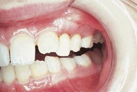
A 9-year-old male visited a dentists office for evaluation of bleeding gums.
Joen Iannucci Haring
History
The patient`s mother stated that the bleeding gums started several months earlier. The area involved included the left maxillary molar region. When questioned about other symptoms, the patient denied any pain in the area. No constitutional symptoms were identified. The patient`s medical history was reviewed. No significant findings were noted.
Examinations
Examination of the head and neck regions revealed no abnormalities present. All vital signs were within normal limits. Oral examination revealed shiny gingiva with a lack of stippling in the area of involvement (see photo). A panoramic radiograph revealed significant bone loss around the maxillary left primary molars (see radiograph).
Differential diagnosis
Based on the clinical information provided, which one of the following is the most likely diagnosis?
- Acute necrotizing ulcerative gingivitis
- Hyperplastic gingivitis
- Gingival fibromatosis
- Early-onset periodontitis
- Granulomatous gingivitis
Diagnosis
Early-onset periodontitis
Discussion
Although periodontitis is typically seen in adults, it may also affect children and adolescents. Early-onset periodontitis is a disorder that is seen in otherwise healthy children, adolescents, and young adults. The three forms of early-onset periodontitis are:
* Prepubertal periodontitis
* Juvenile periodontitis
* Rapidly progressive periodontitis
In contrast to adult periodontitis, early-onset periodontitis is often correlated to one or more deficiencies in the immune response rather than local factors (plaque and calculus, for example). Suspected pathogens for early-onset periodontitis include Actinobacillus actinomycetemcomitans, Prevotella intermedia, and Porphyromonas gingivalis. It is important to note that early-onset periodontitis is not associated with systemic diseases that are known for premature attachment loss (blood dyscrasias, diabetes, Langerhans cell disease, AIDS, hypophosphatasia, for example). And, in fact, such diseases must be ruled out before a definitive diagnosis of early-onset periodontitis is established.
Clinical features
Prepubertal periodontitis is an extremely rare condition seen in younger children; some cases are noted as early as the age of four. Females are affected more frequently than males. Minimal plaque and gingivitis are clinically evident. The presentation is usually localized, and no particular area is involved more than other areas. Prepubertal periodontitis may progress on to become juvenile periodontitis.
Juvenile periodontitis, or localized juvenile periodontitis, is seen in adolescents and young adults under the age of 25. Most cases occur between the ages of 10 and 15. Clinically, a lack of gingival inflammation and a minimal amount of irritants (plaque and calculus, for example) in the presence of deep periodontal pockets are noted. The localized distribution of bone loss is characteristic. The permanent first molar and incisor areas are most often involved. It is believed that this form of periodontitis involves the incisors and molars because these teeth erupt first and have been in the mouth for the longest period of time.
With localized juvenile periodontitis, bilaterally symmetric patterns of bone loss may be seen. The rate of bone destruction is rapid and occurs three to five times faster than that seen in adult periodontitis. The bone loss continues until the teeth are exfoliated, treated, or extracted. Radiographs reveal a vertical pattern of bone loss in the molar and incisor regions. An arc-shaped pattern of bone loss may be noted between the second premolar and second molar, as well as in the anterior regions. Tooth mobility and drifting are common.
When untreated, localized juvenile periodontitis may involve more areas and become generalized. In the generalized form, more teeth are involved, and the bone loss is not restricted to specific areas of the jaws. Most cases of generalized juvenile periodontitis occur between the ages of 12 and 32. As with the localized form of this disease, minimal local irritants are present.
Rapidly progressive periodontitis is a highly destructive form of early-onset periodontitis that is seen in adults between the ages of 20 and 35. The destruction of attachment is rapid and widespread. Alveolar bone loss may take place within several weeks or months, and results in early tooth loss. Clinical features include inflamed and hemorrhagic gingiva. Malaise and weight loss may also be noted.
Treatment
The progression of early-onset periodontitis is not slowed by scaling and root planing. Antibiotics are necessary to eliminate the causative organisms. Such antibiotics (tetracycline, amoxicillin or metronidazole) are recommended along with the mechanical removal of irritants. Periodontal surgery is also recommended.
Antimicrobial rinses are used following surgery. Long-term follow up is required to evaluate for reinfection or incomplete elimination of causative organisms.
Joen Iannucci Haring, DDS, MS, is an associate professor of clinical dentistry, Section of Primary Care, The Ohio State University College of Dentistry.

