Evaluation of attached tissue aids in treatment of recession

By accurately assessing attachment loss, the dental team can respond with increased hygiene efforts or gingival grafts.
Timothy J. Hempton, DDS,
Esther Wilkins, RDH, DMD,
Diane Lancaster, RDH
Loss of attachment can reveal itself clinically as either gingival recession, pocket formation, or a combination of both anatomical developments. Root exposure occurs with gingival recession, and, besides the loss of attachment, other problems surface such as thermal sensitivity, root caries, and an unesthetic appearance.
The width and thickness of the periodontium potentially can limit the development of recession. The first parameter, width, is the occlusal apical dimension of the attached gingiva. The second, thickness, is the buccal lingual dimension of the periodontium adjacent to the root surface (Figure 1). Evaluation of these two parameters can determine the potential risk of recession.
The dental hygienists` understanding of the etiology of recession and the complexities of the tissues involved can enhance prevention strategies.
Measuring the occlusal apical dimension
Here is a quick way to measure the width or the occlusal apical dimension of the attached gingiva. Place your probe on the outside of the tissue and measure from the gingival margin to the mucogingival junction. Now measure the sulcus or pocket depth (probing depth).
Subtract the probing depth from the outside measurement of the gingiva, and you will have the width of attached gingiva. The width of attached gingiva equals the total vertical measurement of gingiva minus the clinical probing depth.
If the periodontal probe extends directly to the mucogingival junction, no measurement of attached gingiva will be obtained, since the patient obviously has no attached gingiva. When the periodontal probe extends apical to the mucogingival junction, mucogingival involvement is present.
Determination of the buccolingual thickness
No standard method has been adopted to determine the buccolingual measurement. However, clinical observation is useful. Thin periodontal structures can easily be noted and differentiated from thicker ones. Prominent roots can be observed clinically. If the roots are quite prominent, one can see the roots directly below thin mucosal tissues since, adjacent to the mucosa or thin gingiva, no bone is present. In photograph #1, thin tissues can be seen on the facial aspects of teeth #23, 24, and 26. Gingival recession can also be observed on the facial aspect of tooth #25. Thin periodontal tissues in a buccolingual dimension may have predisposed tooth #25 to the development of gingival recession.
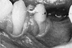
The depth of the dimension includes connective tissue, possibly osseous tissue, and epithelium. The presence of a bony plate will increase this dimension. The osseous crest is typically 1 to 2mm apical to the cementoenamel junction. Two variations of lingual or facial bone position are dehiscences and fenestrations. When the bone dips down more than 2mm from the cementoenamel junction, a dehiscence is formed. In photograph #2, the osseous defect on the buccal aspect of tooth #21 is an example of a dehiscence. If a thinning in the bone creates an aperture or window to the tooth surface, it is a fenestration.
Standard radiographic techniques such as periapical radiographs cannot detect the presence of fenestrations or dehiscences, but computerized tomography may one day provide a cost effective means to do so.
Etiology of recession
Gingival recession is considered to be either trauma-induced or plaque-induced. Examples of trauma-induced recession include:
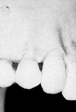
- Toothbrush abrasion. A hard bristle toothbrush or aggressive brushing can result in recession. Gingival tissues can be abraded over a period of time and can lead to root sensitivity. Photograph #3 provides an example. In this case, continued brushing with a hard toothbrush abraded away the gingiva as well as some root structure.
- Self-mutilation. Under certain circumstances, some people traumatize an area as a result of a habit. This trauma may eventually wear away the tissue similar to toothbrush abrasion.
- Surgically induced recession. Gingival recession can be induced via surgical intervention. Examples are gingivectomy procedures (using a scalpel or electrosurgery), modified Widman flap therapy, apically positioned flaps with or without osseous resective therapy, or simply by gingival curettage during phase I therapy.
- Restorative procedures. Preparation for a subgingival crown or a Class V restoration can violate the attachment apparatus. The ensuing chronic inflammation may lead to loss of attachment and subsequent development of gingival recession.
Plaque-induced recession
As mentioned before, the inflammatory response in plaque-induced recession has an impact on both the occlusal apical and buccolingual dimensions. It has been well demonstrated that bacterial plaque is the sole etiologic factor in the initiation of periodontal infections. Although occlusal trauma can be a co-factor, it does not initiate apical migration of the junctional epithelium.
Proper maintenance by the patient and the dental team is of paramount importance in the prevention of the problem. Although oral hygiene and frequent recalls are extremely beneficial, it is important to acknowledge that their effectiveness is limited by broken recall appointments and poor patient habits - a reality of clinical practice. Aware that plaque-induced inflammatory responses can result in continued periodontal breakdown, clinicians must look for other options, including gingival augmentation which replaces mucosal tissue with connective tissue.
Fiber barrier principle
Gingival connective tissue provides a strong defense against inflammation as explained by the fiber barrier principle. This theory has been proposed to explain the protective capabilities of gingival connective tissues as opposed to alveolar mucosal connective tissues. According to the fiber barrier principle, gingival connective tissues, which have concentrated amounts of collagen fibers, may help to reduce the destruction of tissue brought on by the plaque-induced inflammatory process. In-flammation travels along the perivascular spaces. If an area`s vascularity can be minimized, then the inflammatory response will be reduced. If the flow of fluids through the affected surrounding connective tissues can be reduced, then the flow of enzymes de-rived from the inflammatory response will be minimized.
Gingival connective tissues have heavy concentrations of matted collagen fibers. The fiber arrangement can limit the flow of fluids through the involved tissues. In contrast, the alveolar mucosa has a high level of vascularity and very loose connective tissues.
The histological characteristics of attached gingival connective tissue may contain the plaque-induced inflammatory reaction, while alveolar mucosal connective tissue may facilitate its spread. Proponents of the fiber barrier principle advocate a widened band of attached gingival tissue, as opposed to a narrow band of mucosal tissues adjacent to the base of the sulcus. The practice of surgically exchanging alveolar mucosa for grafted gingival tissue is based on the fiber barrier principle.
The measurement of the width of the attached gingival tissue becomes relevant as the junctional epithelium migrates apically, resulting in deeper probing depths. As the base of the sulcus or pocket extends apically, a change in the character of the soft tissue adjacent to the base is significant.
As probing depths increase, the mucogingival junction may be within close proximity to the base of the sulcus or pocket. With increased attachment loss, the base of the pocket may eventually extend beyond the mucogingival junction, creating mucogingival involvement. In this case, mucosal tissues are now adjacent to the root surface at the base of the sulcus or pocket.
As previously mentioned, mucosal tissue has increased vascularity and a loose arrangement of alveolar connective tissue fibers. The soft tissue response to the plaque-induced inflammatory challenge may not be one of containment but rather a magnification of the inflammatory response.
With an increased presence of the cellular and non-cellular components of the immune system present, further loss of attachment and possible tissue necrosis may become evident. The tissue necrosis would be observed as recession.
Clinically, it is common to see mucosa becoming more inflamed in the presence of plaque than gingival tissues. Patients often may experience difficulty brushing areas where there is no zone of attached gingival tissue. Inflamed mucosal tissues can result in significant discomfort, inhibiting patients from proper maintenance of the area. In photograph #2 the character of the soft tissue on the facial aspect of tooth #25 presents a case demonstrating this situation. A 25-year-old patient presented with significant discomfort, no attached gingival tissue, and inflamed mucosa. The patient reported great difficulty maintaining the area.
Influence of the buccolingual thickness
As anatomical features of the width of the attached gingiva influence the development of recession, so does the local anatomy relative to the buccolingual length of the periodontium adjacent to the root surface.
The thickness of buccolingual tissue adjacent to the root surface at the level of the osseous crest (or apical to that level) consists of epithelium, connective tissue, bone, and the periodontal ligament. These tissues vary in thickness.
Increased thickness in this area will reduce the risk for development of recession. The presence of bone will increase this value and decrease the risk of root exposure. In effect, when bone is not present in its normal anatomical position (that is, when a dehiscence or fenestration is present), the soft tissues may be more susceptible to necrosis and, subsequently, recession.
Figure 1 depicts a normal anatomical buccolingual cross section of the hard and soft tissues adjacent to the tooth. Recession should be considered an analog to the development of a periodontal pocket, since both occurrences result in the loss of attachment. Each pathological event is initiated when plaque-induced inflammation results in the invagination of the sulcular epithelium into the underlying connective tissues. Apical migration of the junctional epithelium follows.
The ingress of the epithelium into the underlying connective tissue will increase and the inflammatory process continues. The blood supply to the epithelial tissues comes from the underlying connective tissue. When the periodontal structures are narrow in a buccolingual dimension, the invagination of the epithelium will result in a reduction of the blood supply to both the connective tissue and the epithelium. The decrease in blood supply may lead to necrosis of the affected tissue coronal to the osseous crest and will become evident clinically as recession.
If the attachment apparatus or dentogingival junction is adjacent to a broader length of connective tissue, then the invagination of the epithelium will not likely cut off the connective tissue blood supply. Tissue necrosis will not ensue.
If apical migration of the junctional epithelium continues, however, a pocket will result. To reiterate, loss of attachment will have occurred, but it will be manifested as pocket formation and not as gingival recession.
Gingival augmentation
Various studies have concluded that gingival recession does not always progress, even with inadequate zones of attached gingiva or mucogingival involvement. With the presence of good oral hygiene, recession is unlikely. Patients with less than ideal plaque control, however, may develop gingival recession. To reduce this risk, therapists may opt to increase the zone of attached gingiva.
The surgical techniques during the 1960s and 1970s focused on widening the zone of attached keratinized tissue and included sliding pedical grafts, double papilla grafts, and the free gingival graft. In the 1980s, grafting of connective tissue was introduced.
When a gingival graft procedure is performed, complete healing will occur in approximately 28 days, resulting in a widened band of attached keratinized gingival tissue. Clinically, the tissue is pink in color and may appear lighter than neighboring gingival tissues. The grafted tissue has dense, matted collagen fibers and minimal vascularity. Such microscopic features may be protective in nature and not readily subject to inflammation.
Photograph #4 is the same patient seen in photograph #2, six months after placement of a free gingival graft.
Prevention of attachment loss is a high priority for the dental team. Effective disease control and maintenance by both the dental hygienist and the patient are critical to achieving this goal. Thorough debridement, as well as a review of oral hygiene procedures, are essential components of the dental hygiene visit. Understanding gingival recession aids the clinician in evaluation and monitoring of patients. When problems are recognized as they develop, the dental hygienist is enabled to advise patients about the most beneficial preventive and therapeutic modalities.
Timothy J. Hempton, DDS, and Esther Wilkins, RDH, DMD, are members of the department of periodontology at Tufts University School of Dental Medicine. Diane Lancaster, RDH, is a practicing hygienist in Dedham, Mass.
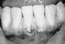
Photograph 1: Gingival recession, tooth #25
Photograph 3: Toothbrush abrasion
Photograph 2: Dehisence, tooth #21
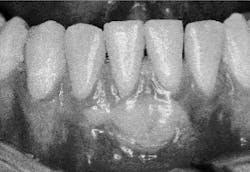
Photograph 4: Gingival augmentation, tooth #25