Case #7
A 67-year-old male visited a dentist for a check-up. Radiographic examination revealed two small round radiopacities attached to the roots of the right maxillary molars.
History

The patient denied any history of signs or symptoms associated with the right maxillary molar region and appeared to be in a general good state of health. The patient’s only significant past medical history included a knee replacement surgery. The patient’s past dental history included sporadic dental examinations and routine restorative, periodontal and prosthetic treatment. At the time of the dental appointment, the patient was not taking medications of any kind.
Examinations
The patient’s blood pressure was recorded at 135/80 and all other vital signs were found to be within normal limits. Examination of the head and neck region revealed no enlarged or palpable lymph nodes. No significant extraoral findings were noted. Examination of the oral soft tissues revealed no unusual findings.
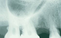
After a thorough clinical examination, periapical radiographs were ordered and exposed of all of the existing teeth in order to evaluate the periodontal condition. Examination of the right maxillary molar radiograph revealed the presence of two small round radiopacities - one attached to the root surface of tooth #2 and one attached to the root surface of tooth #3 (see radiograph). No bony expansion was noted in the area of the involved molars, and no additional radiopacities were noted on the other periapical films.
Clinical diagnosis
Based on the clinical and radiographic information presented, which of the following is the most likely clinical diagnosis?
❏ enamel pearls
❏ calculus
❏ cusps of Carabelli
❏ concrescence
❏ dens invaginatus
Diagnosis
■ enamel pearls
Discussion
Enamel is sometimes found in ectopic locations - on the root of a tooth, for example. The most widely recognized form of enamel in an ectopic location is the enamel pearl (also known as the “enameloma”). This developmental anomaly typically presents as a small globule of enamel attached to a tooth root. As the name suggests, the globule of enamel resembles the shape of a pearl.
The enamel pearl may be comprised of enamel only, or a combination of enamel, dentin, and pulp tissue. The enamel pearl is fairly common. Depending on the population studied, the prevalence of the enamel pearl ranges from 1.1 to 9.7 percent of all patients; Asian populations are most often affected.
Clinical features
Clinically, the enamel pearl appears as a small white dome-shaped mass attached to a root surface. The roots of the maxillary molars are most often involved, while the root surfaces of the mandibular molars are the second most frequent site of involvement. Most enamel pearls develop near the furcation area of a tooth or near the cementoenamel junction.
The enamel pearl may occur singly or in multiples. In most instances, one pearl occurs; however, up to four pearls have been identified on a single tooth. On a dental radiograph, the enamel pearl appears as a small round rediopacity attached to a root surface.
Diagnosis—The diagnosis of an enamel pearl is based on its characteristic radiographic appearance. Occasionally, the enamel pearl may be mistaken for calculus during a clinical exam.
Treatment—If an enamel pearl is detected, the involved root area must be carefully followed (clinically and radiographically) and observed for periodontal attachment problems. Scrupulous oral hygiene must be maintained in an effort to prevent loss of periodontal attachment. In some instances, the removal of the enamel pearl may be desired.
In such cases, if pulp tissue is present within the enamel pearl, a pulp exposure may take place.
Joen Iannucci Haring, DDS, MS, is a professor of clinical dentistry, Section of Primary Care, The Ohio State University College of Dentistry.