Case #2
By Joen Iannucci Haring
A 22-year-old male visited a dental office for a routine checkup. During the examination, a slight, bony swelling was noted near the mandibular first molar. A radiograph of the region revealed a large, round radiopacity.
History
The patient denied any history of pain associated with the mandibular molar area. The patient appeared to be in a general good state of health with no significant medical history. His dental history included routine checkups and restorative dental treatment.
Examinations

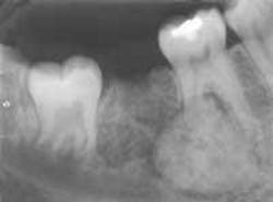
The patient's vital signs were all found to be within normal limits. Extraoral examination of the head and neck region revealed no enlarged or palpable lymph nodes. Intraoral examination revealed a slight bony enlargement of the buccal alveolar bone near the mandibular first molar. Based on this clinical finding, a periapical radiograph of the region was exposed. The film revealed a well-defined, round radiopacity surrounded by a radiolucent rim and obliterating the distal root of the mandibular first molar (see film). The patient was referred to an oral surgeon for biopsy and removal of the lesion.
Clinical diagnosis
Based on the clinical and radiographic information presented, which one of the following is the most likely diagnosis?
• complex odontoma
• periapical cemental dysplasia
• osteoblastoma
• cementoblastoma
• focal sclerosing osteomyelitis
Diagnosis
• cementoblastoma
Discussion
The cementoblastoma is a benign odontogenic tumor that is seen in association with tooth roots. This lesion arises from cementoblasts. The cementoblastoma is not a common lesion and represents less than 1 percent of all odontogenic tumors.
Clinical features
The cementoblastoma is most often seen in the second and third decades of life. More than 50 percent of these lesions are seen in patients under the age of 20. There is no sex predilection. The cementoblastoma is found predominantly in the mandible and affects the posterior region more often than the anterior region. The cementoblastoma is always seen in association with a tooth; the roots of the mandibular first molar are most often affected.
The cementoblastoma typically appears as a slow-growing, solitary lesion. The lesion is associated with a vital tooth. Pain and bony expansion occurs in two-thirds of reported patients. With percussion, there is an audible difference between a tooth with a cementoblastoma and a tooth without a cementoblastoma. Because the tumor is attached to the apex of a tooth, a dull and ankylosed sound is produced when the involved tooth is tapped.
Radiographic features
The cementoblastoma has a fairly characteristic radiographic appearance. The lesion will appear as a well defined radiopaque mass attached to the root of a tooth. The opacity is surrounded by a thin, radiolucent rim. A term that is used to describe the radiographic appearance of a cementoblastoma is a "target" lesion; as the name suggests, the lesion resembles a target.
When viewed on a radiograph, the cementoblastoma typically surrounds the apex of the tooth and extends midway up to the root surface. The cementoblastoma may obliterate the involved root and cause extensive resorption.
In addition, the lesion often appears to be "blended" with the obliterated root surface, and it is difficult to identify where the lesion stops and the root surface begins.
Diagnosis and treatment
A diagnosis of a cementoblastoma cannot be made based on the radiographic appearance alone. A biopsy and microscopic evaluation of the tissue is necessary in order to establish a diagnosis.
The cementoblastoma must be surgically removed along with the involved tooth. If left untreated, the cementoblastoma may continue to grow, destroy bone, and damage adjacent teeth. The cementoblastoma does not recur after total removal.
Joen Iannucci Haring, DDS, MS, is a professor of clinical dentistry, Section of Primary Care, The Ohio State University College of Dentistry.