Case reports: Lasers in soft-tissue management
Case reports: Lasers in soft-tissue management
Jill Taylor, RDH, and
William G. Dickerson, DDS
Lasers have been used in dentistry for the last several years for procedures ranging from soft-tissue cutting to curing and, now, hard-tissue applications. One area under investigation is soft-tissue therapy in the hygiene department. The following is an account of four cases using the Premier Diode Laser (Aurora) for pocket reduction.
Case 1
The first case involves a 64-year-old male with a half-a-pack-a-day smoking habit, but otherwise has a clear health history. This patient had been told that surgery was required, and he was seeking a second opinion on his disease status and options. He had a FMX from September 1997 that showed moderate, generalized bone loss with a vertical distal defect on #20 and multiple crowns and bridges with questionable biological width violations. A full-mouth periodontal probing at his initial appointment recorded generalized 4-6mm pockets with moderate bleeding on probing and mucogingival involvement on #25. Laser therapy with root debridement using microsonics was recommended.
The patient`s second appointment was scheduled for two hours, allowing time to concentrate on #28-#31 and #25-#27. The periodontal etiology was explained to the patient, including the fact that several areas in his mouth were very compromised. Laser therapy was discussed as a conservative way to treat his periodontal disease and that the success of therapy would depend on his contributing factors - i.e., smoking and home therapy. It was recommended that he quit smoking, but the patient refused with the knowledge that perfect tissue most likely would not be achieved due to this refusal.
The patient was prescribed a vitamin therapy of B Complex and C with Bioflavonoids. A local anesthetic was used and reprobing of #25-#31 was performed, noting the furcations. The patient presented 80 percent plaque levels, moderate tobacco stain, and burnished subgingival calculus. Root debridement was completed, first with hand instruments and then with the Dentsply Microsonic scaler. The Premier Diode Laser was used with a 400u fiber at 1.0CW (Continuous Wave) to remove the diseased epithelial lining. The area then was re-lased at 1.8W/10.0 rep rate/0.05 pulse duration to achieve bacterial decontamination. Microsonic irrigation was used with diluted Prosol; applying vitamin E capsules and the recommendation of saline rinses completed the appointment. Six hours of therapy, in total, were completed on the patient with subsequent visits using the same settings on the laser. The administration of a liquid topical Dyclone allowed decontamination on specific sights, retracting (cleaving) the fiber 1-2mm.
The patient was seen six weeks later for his supportive periodontal therapy (S.P.T.) appointment. At this appointment, the recorded pocket depths were reduced from 6-4-6 to 3-2-3. The patient had mostly 2-4mm pockets, with three localized 5mm pockets where vertical bone loss was evident. He still had several areas of 4mm, but the B.O.P. and B.O.S. were slight. It was explained to the patient that his progress would be monitored and any area that may relapse would be treated at the earliest indication. The patient was pleased with the results and thrilled at having avoided surgery.
Case 2
Simultaneously, this patient`s 62-year-old wife was having her periodontal disease treated using the laser. She had a clear health history and was taking no medications. Her bone loss was slight and horizontal. The patient had six old PFM crowns; #29 and #30 were fused. At her initial periodontal therapy appointment, she presented with 4-5mm pockets posterior, 1-3mm anterior, moderate B.O.P., heavy B.O.S., 80 percent plaque, moderate sub- and supra-calculus. She received a total of four hours of laser therapy using 1.0 CW to remove the diseased epithelial lining and then 1.6W/10.0 rep rate/0.05 pulse duration for bacterial decontamination.
When she was seen at her six week S.P.T., her measurements were 2-3mm generalized, no B.O.P. and very slight localized B.O.S. The patient was extremely pleased as she had always bled at her hygiene appointments previous to therapy and she now knew how to floss interproximally #29 and #30.
Case 3
The third case deals with a 78-year-old female. She has a clear health history and takes no medications. She has severe bone loss on all 21 existing teeth; extensive decay on #2 and #3; 4-8mm pockets generalized; 90 percent plaque; black burnished subgingival calculus sporadic; red, edematous, smooth, bulbous papilla; and profuse B.O.P. and B.O.S. She was scheduled for one hour of initial periodontal therapy, six hours on laser therapy and a one hour, six-week S.P.T.
Power was set at 1.4CW, then 1.6W/10.0 rep./0.05 duration, Total Jules 519.1 on her first therapy appointment. On her subsequent visits it went down to 1.2CW and then 1.0CW as it was discovered that the higher wattage was causing the tissue to char, resulting in the possibility of developing scar tissue and not achieving reattachment. The final two hours of therapy were full-mouth bacterial decontamination at 2.0W/10.0 rep./0.05 duration. This is where the most healing took place and the tissue response was excellent.
When the patient was seen at her six-week S.P.T. she had only six sights remaining at 4-6mm. She was re-lased at 1.8W/10.0 rep/0.05 duration. Four of the remaining sights were on teeth #2 and #3, both of which had severe decay below the gumline, close to the bone and had questionable prognosis. Her probe readings on #25 and #26 buccally went from 3-6-7 and 6-4-3 to 2-2-3 and 2-1-2.
Case 4
The fourth case involves the mother of Jill Taylor, one of the authors, who usually is 1-3mm generalized. She experienced a stressful three-month interval and came in with a 6mm pocket on the direct lingual of #12 and several localized 4mm pockets. Jill wanted the pockets reduced to 3mm. Power was set at 1.0CW then 1.6W/10.0 rep./0.05 duration, 935.6 TJ (Total Jules). It is believed that this was far too much energy, even if laser therapy was performed on her full mouth. On the next visit, the power was set at the same for bacterial decontamination, but only 313.8 TJ was used. Less energy was used her next four therapy appointments. When treated at her six-week S.P.T., the power used was only 26.85 TJ in her remaining bleeding points and around #12. Her tissue response was excellent! She is back to 1-3mm with no B.O.P. and slight B.O.S.
It is evident that this new technology is an exciting therapeutic option to offer patients. There is a learning curve with this technique and it may be possible to achieve better results using less wattage. Use of the laser for periodontal therapy appointments has allowed patients to gain tissue reattachment and improved texture that was not previously available from conventional conservative treatment. It is important also to understand the necessity to charge for such treatment. The laser is an expensive tool and one must achieve a return on the investment. The benefit to the patient is that the cost of therapy is considerably less than surgery.
Jill Taylor, RDH, has worked in cosmetic-oriented practices for more than 12 years. She is a clinical instructor for JP Consultants. Ms. Taylor also consults with dental manufacturers on new product developments and product enhancements. She currently practices in Las Vegas, Nevada. Dr. William Dickerson lectures internationally on esthetic dentistry and marketing a cosmetic practice. He is visiting clinical professor and director of the Comprehensive Esthetic Restorative Continuum at Baylor College of Dentistry, as well as director of the Las Vegas Institute for Advanced Dental Studies. He has had more than 100 articles published in national dental journals on techniques and innovative uses for esthetic restorations, as well as marketing and managing a cosmetic dental practice. Dr. Dickerson is past editor of the AACD Journal. He maintains a cosmetic-oriented restorative practice in Las Vegas, Nev.
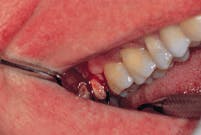
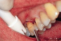
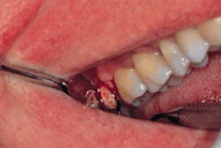
In the top photograph, a diseased area of infection is shown. In the middle photo, 1.0 CW removes the diseased epithelial lining. At the six-week recall, the bacterial decontamination (bottom photo) is countered with 1.6W, 10.0 rep rate, and .05 pulse.