Oral Exam: Mesiodens
By Nancy W. Burkhart, BSDH, EDD
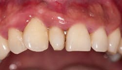
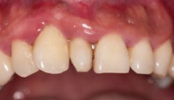
The image depicted here is one of what is termed a supernumerary tooth. In this case, it is called a mesiodens (see Figure 1). Supernumerary teeth are in excess of the normal number of teeth, and they are further classified according to their location.
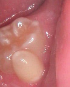
A mesiodens is a supernumerary tooth in the maxillary anterior incisor region. If the supernumerary tooth were found in the posterior region it would be termed a distomolar or distodens (4th molar). If the tooth happens to be found lingually or buccally, the term paramolar is used (see Figure 2). Developmental alterations can occur affecting the number of teeth, the size of teeth, the shape of teeth, and the structure of teeth.
---------------------------------------------------------------------------
Other articles by Burkhart
---------------------------------------------------------------------------
Anodontia refers to a lack of tooth development and usually does not occur in the deciduous dentition. The teeth most often affected are the third molars, the second premolars, and the lateral incisors.
Hypodontia refers to the lack of development of one or more teeth. Hyperdontia refers to an increase in the number of teeth and occurs more frequently in the permanent dentition. Most hyperdontia occurs in the maxilla and specifically the anterior region.
Dental transposition refers to normal teeth erupting in an inappropriate position.
Hyperdontia refers to additional teeth as stated above and mesiodens fall into this definition. The number of those affected with hyperdontia is approximately 0.15% to 3.9% of the population. Most supernumerary teeth occur in the maxillary arch, they occur unilaterally, and over half are found in the anterior region. It is not uncommon to find other supernumerary teeth within the same patient, and there may also be other craniofacial anomalies as well. Russell and Folwarczna (2003) distinguished between the types of mesiodentes such as rudimentary (defined as those occurring in the permanent dentition), primary (those occurring as supplementary), and those according to morphology such as conical, tuberculate or molarform.
Etiology: The etiology of mesiodens is unclear, but males are twice as affected as females in permanent teeth (possibly an autosomal recessive gene), and there is a familial trait. The genetic susceptibility along with environmental factors appears to increase the activity of the dental lamina leading to the mesiodens. The hyperactivity theory appears to be the most accepted possibility, although genetic transmission is also favored. An additional theory is that the tooth bud splits, creating two teeth.
Reported increases of mesiodens within various ethnic groups are documented. For instance, supernumerary teeth in Japanese children are reported to be 0.05%, in Canadian pupils, 0.64%, and in Caucasian general populations between 0.1 and 3.8%. Higher percentages are reported in African and Asian populations as well.
The mesiodens interfere with eruption and cause other alignment problems with the existing teeth. Only a small portion of supernumerary teeth eventually erupts (approximately 25%) and the pattern of eruption is usually the first indication of a problem in the case of mesiodens. Because of the concern in eruption patterns, the dental professional usually orders radiographs to assess any problems that may be unseen clinically. Radiographs provide some assistance, but are often vague because of primary teeth obstruction.
Early intervention is suggested to prevent additional damage such as misalignment and delayed eruption of the permanent central incisors. Meighani and Pakdaman (2010) point out in a review of the literature that some practitioners prefer to wait to remove mesiodens until the root of the central and the lateral teeth are completely formed, so that damage to the permanent tooth is not an issue.
Additionally, there is an opinion that the removal in a mixed dentition is best served by removal around age 10 years so that proper alignment occurs and less orthodontic or surgical procedures are needed. Mesiodens are associated with dentigerous cysts that are developmental cysts of odontogenic origin. These types of cysts are found around the crowns of supernumerary teeth and mesiodens.
Mesiodens have been found in certain syndromes such as cleft lip and palate, cleidocranial dysostosis, and Gardner's syndrome. Supernumerary teeth in general have associations with Ehler-Danlos syndrome, Apert syndrome, and Down's syndrome as well.
The concerns associated with mesiodens are delayed eruption of permanent teeth, cyst formation, abnormality of the roots, multilobulated supernumerary teeth, crowding, diastemas, root dilacerations, resorption of the roots of adjacent teeth, and eruption of permanent teeth into the nasal cavity. In a study of 5,000 patients, Kumar and Gopal (2013) found 112 supernumerary teeth in 78 subjects. Twenty-nine teeth were mesiodens, or 25.8% of all supernumerary teeth found within the study.
Conclusions: Removal of the mesiodens is needed because of the above stated reasons, and researchers also point out the fact that the tooth bud displacement impedes oral hygiene and introduces the tooth to a predilection of caries defects and marginal periodontitis. The identification of medsiodens also heightens the awareness of the clinician to further evaluate the patient for other health issues, syndromes associated with mesiodens and additional supernumerary teeth that may be yet undiagnosed within the patient. Radiographs are extremely important in making a definitive diagnosis.
As always, keep listening to your patients and continue to ask good questions.
NANCY W. BURKHART, BSDH, EdD, is an adjunct associate professor in the department of periodontics, Baylor College of Dentistry and the Texas A & M Health Science Center, Dallas. Dr. Burkhart is founder and cohost of the International Oral Lichen Planus Support Group (http://bcdwp.web.tamhsc.edu/iolpdallas/) and coauthor of General and Oral Pathology for the Dental Hygienist. She was a 2006 Crest/ADHA award winner. She is a 2012 Mentor of Distinction through Philips Oral Healthcare and PennWell Corp. Her website for seminars on mucosal diseases, oral cancer, and oral pathology topics is www.nancywburkhart.com.
References
Delong L, Burkhart NW. General and Oral Pathology for the Dental Hygienist. 2nd ed. Wolters Kluwer-Lippincott. Williams & Wilkins. Baltimore 2013.
Burkhart NW. Double teeth leads to differential diagnosis. RDH. 2010; 30(1): 46-47, 67.
Kumar DK, Gopal KS. An epidemiological study on supernumerary teeth: a survey on 5,000 people. J Clinical and Diagnostic Research. 2013; Jul. 7(7): 1504-1507.
Meighani G, Pakdaman A. Diagnosis and management of supernumerary (mesiodens): a review of the literature. Journal of Dentistry, Tehran University of Medical Sciences, Tehran Iran. 2010; 7(1): 41-49.
Russell KA, Fowlwarczna MA. Mesiodens-Diagnosis and management of a common supernumerary tooth. J Can Dent Assoc 2003; 69(6): 362-6.
Past RDH Issues


