The uvula: An unnoticed appendage
Editor's note: Originally published November 8, 2019. Formatting updated September 5, 2022.
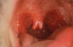
It has been suggested that the uvula is instrumental in directing the mucous secretions that drain from the nasal cavity to the back of the tongue. Other purposes include protection from the eustachian tubes, immunological functions, protection of the throat from hot foods and liquids, and encouraging proper direction of solid foods. We have known about the bifid uvula in connection with cleft palate cases for some time, yet some authors have reported no known functions for the uvula in decades past (figure 1).1,2
Research in the past few years has highlighted the importance of the uvula in speech patterns. The uvula prevents excessive nasality of the voice by controlling resonance of air over the larynx. The partial or total removal of the uvula affects important sounds in French, Arabic, and certain West African languages, as well as in the Yorkshire dialects of the United Kingdom.1,2
The multiple etiologies of angular cheilitis
Lichen planus pemphigoides: An autoimmune blistering disease
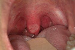
In recent years, the role of the uvula in snoring and in obstructive sleep apnea (OSA) has been a major issue with many studies underway. The uvula consists of connective and granular tissue with diffuse interdigitated muscle fibers.2 People with OSA have thicker, larger uvulas with more muscle and fat.2 A review by Chang et al. states that histologic analysis has shown people with OSA to have bulkier, more edematous uvulas with thicker epithelia and increased leukocytes when compared to samples from controls with no history of snoring.3 The authors question whether the increase in size or epithelium is due to the more rigorous motion experienced during snoring (figure 2).
Uvulectomy, or removal of the uvula (figure 3), has been discussed in writings dating back to AD 324–1453, and it has been equated with the cure of disease since ancient times.2 In some countries, it is a ritual to remove uvulas from toddlers or infants of both sexes. In early years, the uvula was removed with the tonsils because of the possibility of infections. In many countries, particularly in the Middle East and Africa, uvulectomy has been considered a way to keep the person healthy. In certain cultures, the uvula is removed because people believe that uvulectomy cures disease, encourages better breastfeeding, or has other curative effects.1,4 Uvulas that appear to be malformed are often removed as well. In Nigeria and Sudan, elongated uvulas are often surgically removed due to a belief that malformed uvulas promote lifelong throat problems.4,5 Traditional healers, or mgangas, in Tanzania are reported to remove elongated uvulas in children and babies due to chronic coughing, generalized malaise, and weight loss issues.1 Traditional treatment is still used for other health issues in most cases, but the removal of the uvula produces the most serious consequences due to severe bleeding after removal. Usually no anesthesia is administered, and the disinfection techniques can be suspect. The practice of removing canines for treatment of vomiting and diarrhea is also practiced today in smaller numbers of cases at this time.5
Interestingly, the uvula secretes several types of thin saliva comprising both serous and mucous components. The secretion is mostly fluid and lubricates the postpharyngeal areas, released when needed by muscle contraction.6 Most of the soft palate has mucous glands that produce a thicker type of secretion. The uvula has the ability to move backward and forward during certain functions, releasing the fluid when needed and basting the pharynx with lubrication. This lubrication is known to aid in speech by lubricating the vocal cords. Patients who have their uvulas removed are more prone to dryness in the posterior of the mouth.2
The removal of the uvula has been a form of treatment for snoring and obstructive sleep apnea for some time. Recent reviews in the literature have highlighted the implication of neurogenic pathology underlying the loss of soft palate and uvular tone in the progression of snoring and sleep apnea.7 The relationship between sleep-disordered breathing and the size, length, and width of the uvula lacks data. In a recent systematic review, authors suggested a direct correlation between uvula size and snoring, with larger uvulas correlating with more severe snoring and obstructive sleep apnea. The findings noted a uvular length of greater than 15 mm was considered elongated and a uvular width greater than 10 mm was considered wide. The authors suggest more research in snoring and OSA for the future.3
Bifid uvulas are documented as observed anomalies and are thought to be the minimum evidence of cleft palate occurrence in early formation. According to Neville et al., the prevalence of cleft uvula is much higher than that of cleft palate, affecting one in 80 white individuals. The frequency in Asian and Native American populations is as high as 1 in 10. Cleft uvula is less common in black people, occurring in one out of every 250 people.8 Submucous palatal cleft is noted along with the bifid uvula in some cases. This is a defect in the musculature of the soft palate and may appear as a bluish midline discoloration.9
Some techniques that alter the uvula, such as radiofrequency ablation, cause the uvula to heal with scarring and then become less mobile. Because of the lubricating qualities of the uvula, many patients who have had a uvulopalatoplasty may complain of dryness in the posterior/throat areas. Recommending products and procedures to relieve xerostomia assists the patient in reducing this discomfort.7
Many offices are becoming trained in detecting problems related to OSA and have become well versed in conversing and recommending treatment or referral when complaints are voiced by patients related to sleep issues and xerostomia. Noting conversations, documenting findings, and taking oral images assists in evaluating complaints and changes in the posterior pharyngeal area. The uvula is important in the long-term care of patients. When findings appear to require a referral to someone who specializes in OSA, it is of great benefit to the patient (or the patient’s parent) that complete records are sent to the specialist or those rendering treatment.
With a more diverse population, there is a great chance you will see someone who has had his or her uvula removed, either through treatment for snoring or perhaps through ritual practices. There is a very likely chance that you will also notice a variation in the shape, size, movement, or texture of the appendage, as well, when you are paying attention to the uvula (figure 4).
As always, continue to ask good questions and always listen to your patients.
References
1. Manni JJ. Uvulectomy, a traditional surgical procedure in Tanzania. Ann Trop Med Path. 1984;78:49-53. 2. Back GW, Nadig S, Uppal S, Coatesworth AP. Why do we have a uvula?: Literature review and a new theory. Clin Otolaryngol Allied Sci. 2004;29(6):689-693.3. Chang ET, Baik G, Torre C, Brietzke SE, Camacho M. The relationship of the uvula with snoring and obstructive sleep apnea: a systematic review. Sleep Breath. 2018;22(4):955-961. doi:10.1007/s11325-018-1651-5.4. Ijaduola GTA. Uvulectomy in Nigeria. J Laryngol Otol. 1981;95(11):1127-1133. doi:10.1017/S0022215100091908.5. Satti SA, Mohamed-Omer SF, Hajabubker MA. Pattern and determinants of use of traditional treatments in children attending Gaafar Ibnauf Children’s Hospital, Sudan. Sudan J Paediatr. 2016;16(2):45-50.6. Finkelstein Y, Meshorer A, Talmi YP, Zohar Y, Brenner J, Gal R. The riddle of the uvula. Otolaryngol Head Neck Surg. 1992;107(3):444-450. 7. Patel JA, Ray BJ, Fernandez-Salvador C, Gouveia C, Zaghi S, Camacho M. Neuromuscular function of the soft palate and uvula in snoring and obstructive sleep apnea: A systemic review. Am J Otolaryngol. 2018;39(3):327-337.8. Neville BW, Damm DD, Allen CM, Chi AC. Oral and Maxillofacial Pathology. 4th ed. St. Louis, MO: Elsevier; 2016. 9. Sales SAG, Santos ML, Machado RA, et al. Incidence of bifid uvula and its relationship to submucous cleft palate and a family history of oral cleft in the Brazilian population. Braz J Otorhinolaryngol. 2018;84(6):687-690.About the Author
Nancy W. Burkhart, EdD, MEd, BSDH, AAFAAOM
NANCY W. BURKHART, EdD, MEd, BSDH, AAFAAOM, is an adjunct professor in the Department of Periodontics-Stomatology, College of Dentistry, Texas A&M University, Dallas, Texas. She is founder and cohost of the International Oral Lichen Planus Support Group (dentistry.tamhsc.edu/olp/) and coauthor of General and Oral Pathology for the Dental Hygienist, now in its third edition. Dr. Burkhart is an academic affiliate fellow with the American Academy of Oral Medicine, where she also serves as chair of the Affiliate Fellowship Program Committee. She was awarded the Dental Professional of the Year in 2017 through the International Pemphigus and Pemphigoid Foundation, and she is a 2017 Sunstar/RDH Award of Distinction recipient. Her professional interests are in the areas of oral medicine and the relationship between oral and whole-body health, with a focus on mucosal disease and early oral cancer detection. Contact her at [email protected].


