The forgotten ulcer: When herpesviruses reveal themselves in the dental chair—connecting HSV, CMV, and EBV in transplant patients
Key Highlights
- Persistent, painless oral ulcers in transplant patients may signal viral reactivation (especially CMV)—not trauma—requiring timely biopsy and medical collaboration.
- HSV, CMV, and EBV share a latent herpesvirus biology, with immunosuppression allowing atypical, overlapping, and more aggressive oral presentations.
- Missed diagnoses often stem from pattern recognition without context—medical history, lesion behavior, and interdisciplinary communication are critical for early detection.
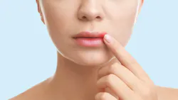
A 47-year-old male presents for a routine recare appointment. His medical history notes a kidney transplant eight months ago. As you begin the appointment, he casually mentions a sore in his mouth that has been there for a few weeks. It does not seem to be improving. It does not particularly hurt. He assumed it would resolve on its own.
On examination, you see an ulcer on the soft palate. It is deep, with irregular borders and a slightly necrotic appearance. It does not immediately fit into a familiar category, but it also does not feel urgent at first glance.
You review the previous note from three months prior. The lesion had already been present. It was documented as likely traumatic. No referral was placed.
Weeks later, after evaluation by his transplant team, the diagnosis becomes clear—cytomegalovirus (CMV)-associated oral ulceration, a reactivated herpesvirus, taking advantage of an immunosuppressed host … a lesion that had been visible long before it became part of a systemic picture.
What was missing was not the finding; it was the connection
In clinical practice, we are trained to recognize patterns. A small ulcer is often traumatic. A recurrent vesicular lesion suggests herpes simplex virus (HSV). A white corrugated plaque on the lateral tongue may point toward Epstein-Barr virus (EBV).1 Each lesion is categorized, labeled, and managed within its own diagnostic box.
But in transplant patients, this way of thinking can be limiting, because what we are often seeing is not a single disease process, but different clinical expressions of the same viral family (figure 1).
A shared biology: The herpesvirus family
HSV-1, HSV-2, CMV, and EBV all belong to the herpesvirus family. What unites them is not just their structure, but their behavior.2
Once acquired, these viruses are never eliminated. They establish latency within the body and remain under immune control, often for life. In immunocompetent individuals, this balance is stable; HSV may reactivate intermittently as herpes labialis, while CMV and EBV typically remain silent.
Additional reading: Why viruses should be part of the health history
What we need to understand is that transplant patients exist in a different biological context. Immunosuppressive therapies, essential for graft survival, disrupt the mechanisms that keep these viruses contained. And what follows is not simply reactivation, but a shift in how these viruses behave clinically. HSV lesions may become more extensive or atypical. CMV may manifest as large, nonhealing ulcers. EBV may present as oral hairy leukoplakia or, in more severe cases, as part of a lymphoproliferative process.3
The key is not the individual virus; it is the loss of immune control over a family of latent viruses. And in many cases, the oral cavity is where that loss first becomes visible.
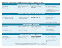
The immunosuppressive regimen and the viral opportunity
Transplant recipients do not take a single medication. They take a combination of agents, each targeting a different arm of the immune response, and that layered approach creates a compounded vulnerability to viral reactivation.
Calcineurin inhibitors: Silencing the T-cell response
Tacrolimus and cyclosporine block calcineurin, an enzyme essential to T-cell activation, preventing the production of interleukin-2 and shutting down adaptive immune surveillance. The consequence for latent herpesviruses is direct: without CD8+ T-cells actively containing them, CMV and EBV do not need a trigger to reactivate. They only need the absence of control.4
Antimetabolites: Depleting the immune repertoire
Mycophenolate and azathioprine reduce both T and B lymphocyte populations by inhibiting purine synthesis. For EBV specifically, this is a double loss; B lymphocytes are both the primary reservoir of latent virus and the source of the antibody response that limits its spread. When their numbers fall, the containment falls with them.5
Corticosteroids: Broad suppression at every level
Prednisone suppresses innate and adaptive immunity simultaneously, impairing cytokine production, macrophage function, and mucosal defenses, including those of the oral cavity. It creates the conditions where HSV, CMV, and EBV can reactivate concurrently, and where a single reactivation event goes uncontrolled far longer than it would in an immunocompetent host.6
mTOR inhibitors: A different risk profile
Sirolimus and everolimus carry a lower viral risk than calcineurin
inhibitors—but they introduce a distinct oral complication: aphthouslike ulcers that closely mimic viral lesions. A nonhealing ulcer in a patient on sirolimus may represent drug toxicity, viral infection, or both. That distinction requires laboratory confirmation, not clinical observation alone.7
CMV: The underestimated oral pathogen
CMV is one of the most common opportunistic infections in transplant medicine, yet its oral manifestations are still frequently overlooked.
Clinically, CMV-associated ulcers tend to present as larger, deeper lesions with irregular borders. They may have a necrotic appearance and can occur on any mucosal surface, including areas we do not typically associate with aphthous ulcers, such as the hard palate or attached gingiva. What makes them particularly deceptive is not only their appearance, but their behavior: they are slow to heal or do not heal at all, and they often present with surprisingly little pain relative to their size.8
And that absence of pain matters. Pain is often what drives both the patient’s concern and the clinician’s urgency. When it is missing, the lesion feels less urgent. The follow-up becomes less immediate. Time passes.
The diagnosis requires biopsy and laboratory confirmation. But before that step ever happens, there has to be a moment of recognition, a moment where something does not fully align with the expected clinical picture.
EBV: More than what we learned in school
EBV is familiar to most clinicians in its classical forms: infectious mononucleosis and oral hairy leukoplakia. But in transplant patients, its clinical significance extends further.
One of the most important associations is post-transplant lymphoproliferative disorder (PTLD9), a spectrum of conditions ranging from relatively benign proliferations to aggressive lymphomas. In some cases, the oral cavity is one of the first places where these changes manifest: painless swellings, areas of ulceration, or exophytic lesions that can easily be mistaken for reactive or odontogenic processes.10,11 What makes them challenging is not their rarity, but their familiarity. They resemble things we see every day.
Oral hairy leukoplakia—the corrugated, nonwipeable white lesion typically found on the lateral tongue—is not just an incidental finding in an immunosuppressed patient. It is a reflection of EBV activity and, more importantly, of the level of immune suppression the patient is experiencing.12,13
Why these lesions are often missed
One of the greatest challenges with these lesions is how ordinary they can appear. They resemble aphthous ulcers. They resemble trauma. They fit patterns that feel familiar, and therefore safe.
But in oral pathology, clinical reasoning is not just about what a lesion looks like; it is about the context in which it appears. An ulcer that persists beyond two weeks in a transplant patient is not the same clinical entity as that same ulcer in a healthy individual. Location, behavior, and healing (or the lack of it), matter. And above all, the patient’s systemic condition matters.
When these findings are overlooked, it is rarely due to a lack of knowledge alone. More often, it is the result of small gaps aligning: incomplete medical histories, patients who do not recognize the relevance of their transplant to an oral finding, pattern recognition working against us when the clinical context shifts, and the absence of pain, creating a false sense of reassurance.
But perhaps the most important gap is structural. Medical and dental care often function in parallel rather than in collaboration. The transplant team manages systemic risk; the dental team observes the oral environment. Without communication between them, both perspectives remain incomplete, and the lesion that connects them goes unrecognized.
The medications that preserve a transplanted organ dismantle, layer by layer, the immune architecture that would otherwise destroy it. This dismantling creates the conditions in which viruses that have coexisted silently for decades are suddenly free to move. The oral cavity is often where that movement begins, and the clinician who recognizes it is the one who can act before that window closes.
Editor's note: The article appeared in the June 2026 print edition of RDH magazine. Dental hygienists in North America are eligible for a complimentary print subscription. Sign up here.
References
- Shahrabi-Farahani S, Aguirre S. Herpesvirus-related lesions of the oral mucosa. Oral Maxillofac Surg Clin North Am. 2023;35(2):175-187. doi:10.1016/j.coms.2022.10.012
- Lee DH, Zuckerman RA, AST Infectious Diseases Community of Practice. Herpes simplex virus infections in solid organ transplantation: guidelines from the American Society of Transplantation Infectious Diseases Community of Practice. Clin Transplant. 2019;33(9):e13526. doi:10.1111/ctr.13526
- Mainville GN, Marsh WL, Allen CM. Oral ulceration associated with concurrent herpes simplex virus, cytomegalovirus, and Epstein-Barr virus infection in an immunocompromised patient. Oral Surg Oral Med Oral Pathol Oral Radiol. 2015;119(6):e306-e314. doi:10.1016/j.oooo.2014.10.019
- Lam E, Bashir B, Chaballa M, Kraft WK. Drug interactions between direct-acting oral anticoagulants and calcineurin inhibitors during solid organ transplantation: considerations for therapy. Expert Rev Clin Pharmacol. 2019;12(8):781-790. doi:10.1080/17512433.2019.1637733
- Nayagam JS, Heneghan MA, Samyn M, Joshi D. Epstein-Barr virus status and immunosuppression use in paediatric autoimmune liver disease. Aliment Pharmacol Ther. 2022;55(4):455-463. doi:10.1111/apt.16708
- Razonable RR. Management strategies for cytomegalovirus infection and disease in solid organ transplant recipients. Infect Dis Clin North Am. 2013;27(2):317-342. doi:10.1016/j.idc.2013.02.005
- Sibaud V, Boralevi F, Vigarios E, Fricain JC. Toxicité endobuccale des thérapies ciblées anticancéreuses [Oral toxicity of targeted anticancer therapies]. Ann Dermatol Venereol. 2014;141(5):354-363. doi:10.1016/j.annder.2014.03.009
- Olczak-Kowalczyk D, Pawłowska J, Cukrowska B, et al. Local presence of cytomegalovirus and Candida species vs oral lesions in liver and kidney transplant recipients. Ann Transplant. 2008;13(4):28-33.
- Krasuska-Sławińska E, Minko-Chojnowska I, Pawłowska J, Dembowska-Bagińska B, Pronicki M, Olczak-Kowalczyk D. Post-transplant lymphoproliferative disorder (PTLD) manifesting in the oral cavity of a 13-year-old liver transplant recipient (LTx). Ann Transplant. 2015;20:478-482. doi:10.12659/AOT.893497
- Johnson J, Kerecuk L, Harrison M, Taylor JO, Odell E. Epstein-Barr virus-associated lymphoproliferative disease in oral cavity in a renal transplant recipient: a case report. Pediatr Transplant. 2007;11(3):340-344. doi:10.1111/j.1399-3046.2007.00694.x
- Akbas A, Tiede C, Lemound J, Maecker-Kolhoff B, Kreipe H, Hussein K. Post-transplant lymphoproliferative disorders with naso- and oropharyngeal manifestation. Transplant Int. 2015;28(11): 1299-1307. doi:10.1111/tri.12632
- Alramadhan SA, Bhattacharyya I, Cohen DM, Islam MN. Oral hairy leukoplakia in immunocompetent patients revisited with literature review. Head Neck Pathol. 2021;15(3):989-993. doi:10.1007/s12105-021-01287-8
- Almazyad A, Alabdulaaly L, Noonan V, Woo SB. Oral hairy leukoplakia: a series of 45 cases in immunocompetent patients. Oral Surg Oral Med Oral Pathol Oral Radiol. 2021;132(2):210-216. doi:10.1016/j.oooo.2021.03.015
About the Author

Andreina Sucre, MSc, RDH
Andreina Sucre, MSc, RDH, is an international dentist, oral pathology, and oral surgery specialist practicing dental hygiene in Miami, Florida. A passionate advocate for early pathological diagnosis, she empowers colleagues through lectures focused on oral pathologies. Andreina is the founder of The Patho RDH, a published author for RDH magazine, and a selected speaker at RDH Under One Roof 2026. Committed to community outreach, she educates non-native English-speaking children on oral health and actively volunteers in dental initiatives.