Gingipains: Exploring their impact on overall health beyond periodontal disease
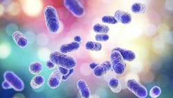
Porphyromonas gingivalis (Pg), a key pathogen in periodontitis, possesses a range of virulence factors that aid its colonization in the gingival sulcus and lead to the onset of disease. Virulence factors of Pg encompass polysaccharide capsules, lipopolysaccharides, fimbriae, hemagglutinins, vesicles, outer membrane proteins, IgA and IgG proteases, and cysteine proteinases along with their associated adhesins. These factors engage in diverse activities within the host, including tissue destruction, perturbation of host defenses, and bone resorption.1,2
Pg releases outer membrane vesicles (OMVs) containing gingipains, which are vital enzymes in its pathogenicity. OMVs, small spherical structures, are shed from the outer membrane of gram-negative bacteria such as Pg, carrying various bacterial components such as lipids, proteins, and nucleic acids, including gingipains. Gingipains play a crucial role in Pg’s pathogenicity through both direct and indirect activities and are pivotal in all stages of infection, including attachment, colonization, nutrient acquisition, evasion of host defenses, tissue invasion, and dissemination.3
Gingipains, classified as cysteine proteinases, constitute a crucial family of proteolytic enzymes and are key contributors to the pathogenicity of Pg. These enzymes are grouped into three main types based on their substrate specificity: arginine-specific (RgpA and RgpB) and lysine-specific (Kgp). Importantly, these three cysteine proteinases, along with their associated adhesins, are acknowledged as the primary virulence factors of Pg.4,5 The enzymes are named based on their ability to break down proteins at specific amino acid sites—arginine-specific gingipains target arginine amino acids in proteins, while lysine-specific gingipains target lysine amino acids.
Arginine-specific gingipains (RgpA and RgpB)
RgpA and RgpB have specific cutting abilities, primarily targeting arginine amino acids in proteins. These enzymes are crucial for breaking down host proteins such as extracellular matrix components, cell adhesion molecules, and cytokines, leading to tissue damage in periodontal disease. While some proteins are fully degraded, providing nutrients for Pg, others are only partially broken down, evading the body’s defense mechanisms and hindering Pg clearance. Additionally, RgpA and RgpB can degrade host immunoglobulins, complement proteins, and cytokines, weakening the host immune response and supporting bacterial survival in the periodontal pocket. Moreover, they contribute to the activation of pro-inflammatory signaling pathways in host cells, fueling the chronic inflammatory response characteristic of periodontal disease. Arginine-specific cysteine proteinases can activate the anticoagulant protein C, inhibiting blood coagulation, which could contribute to the bleeding tendency of periodontal sites.6
Lysine-specific gingipain (Kgp)
Kgp is another major protease produced by Pg that specifically cleaves peptide bonds following lysine residues. Like RgpA and RgpB, Kgp is involved in degrading host proteins and evading host immune responses. Kgp has been shown to play a role in tissue destruction and immune modulation in periodontal disease, albeit with some differences in substrate specificity and regulatory mechanisms compared to RgpA and RgpB.
Interconnection of inflammatory processes
Cytokines play a crucial role in regulating the immune response and orchestrating inflammation. Gingipains dysregulate the cytokine network in periodontal disease, leading to chronic inflammation, tissue destruction, and disease progression. They directly stimulate and indirectly activate inflammatory cytokines such as tumor necrosis factor-alpha (TNF-α), interleukin-1 beta (IL-1β), interleukin-6 (IL-6), and interleukin-8 (IL-8), exacerbating periodontal tissue damage.
Furthermore, gingipains influence the expression and activation of host matrix metalloproteinases (MMPs). Kgp activates MMP-1 and MMP-9, while RgpA activates MMP-3. MMPs degrade collagen, a critical component of the extracellular matrix, contributing to the formation of periodontal pockets and progression of periodontal disease.7 Fibrinogen, a soluble glycoprotein found in blood plasma, contributes to the inflammatory response in periodontal disease and can exacerbate tissue damage when deposited in periodontal tissues. Gingipains interact with fibrinogen by cleaving it, disrupting blood clot formation and wound healing. This cleavage releases bioactive fragments, such as fibrinopeptides, which modulate immune responses, exacerbating inflammation and tissue damage. Fibrinogen also serves as a substrate for bacterial adhesion and invasion. By cleaving fibrinogen, gingipains facilitate the adherence of Pg to host tissues, promoting bacterial invasion into the bloodstream and systemic complications associated with periodontal disease.
Systemic complications
Emerging evidence suggests that gingipains may exert effects beyond the oral cavity, potentially contributing to systemic health conditions. For cardiovascular health, gingipains degrade junctional proteins in the oral epithelium, facilitating bacterial translocation and systemic dissemination. They stimulate the production of pro-inflammatory cytokines and chemokines, promoting systemic inflammation and immune cell recruitment. Gingipains impair endothelial function and promote vascular inflammation, contributing to the pathogenesis of cardiovascular disease, and have been detected in atherosclerotic plaques.8 Gingipains activate coagulation cascades and platelet aggregation, potentially increasing the risk of thrombotic events. They are associated with endothelial dysfunction, inflammation, and plaque instability.9
Gingipains affect metabolic health by exacerbating insulin resistance and impair glycemic control through their pro-inflammatory effects and modulation of adipose tissue function. Adipose tissue dysfunction is a hallmark of obesity and insulin resistance, characterized by aberrant adipokine secretion, impaired lipid storage, and inflammation within adipose tissue. When gingipains were translocated to the pancreas in mice, there were significant changes in architecture and B cell apoptosis, which may be involved in the development of prediabetes.10
Gingipains can cleave lipid-binding proteins or modify lipid components, potentially altering host lipid metabolism or signaling pathways. They may also influence the inflammatory response by modulating lipid-derived mediators or lipid metabolism in immune cells.
Gingipains have the potential to induce citrullination of host proteins, initiating autoimmune responses, and play a role in the development of rheumatoid arthritis (RA). Dysregulated citrullination is central to the generation and persistence of antibodies against citrullinated proteins, a defining characteristic of RA. They can contribute to the degradation of collagen and other joint tissues, a hallmark of RA, and can activate immune cells, stimulating the production of inflammatory molecules and thereby intensifying joint inflammation and tissue damage. 11
When tau, a type of protein found in nerve cells in the brain, encounters the gingipains enzyme, it undergoes a transformation. The tau protein is released from the nerve cell and undergoes physical changes, forming coils and noncoiling filaments. These altered forms of tau then reattach to the nerve cell and become part of the lesion known as neurofibrillary tangles, which are a hallmark of Alzheimer’s disease (AD) and ultimately lead to the death of nerve cells. When a nerve cell dies and the tau protein is released into the brain, the tau protein can attach itself to neighboring healthy nerve cells, initiating the same process and causing further damage to the brain as the disease spreads.12 Gingipains released by Pg can also contribute to the formation of amyloid-beta plaques that form on the brains of those suffering from Alzheimer’s.13
Given their pivotal role in the pathogenesis of periodontal disease, gingipains have become promising targets for therapeutic intervention. Various strategies have been investigated to inhibit gingipain activity, including the development of small-molecule inhibitors, antibodies, and vaccines. These approaches aim to disrupt Pg virulence mechanisms, reduce periodontal inflammation, and restore tissue balance in the periodontium. Something as simple as cranberry and rice extracts have also shown potential in interfering with gingipain activity, inhibiting formation and growth of periodontal pathogens. While the ideal gingipain inhibitor remains elusive, targeting gingipains represents a novel approach to managing and preventing periodontitis.
Editor's note: This article appeared in the June 2024 print edition of RDH magazine. Dental hygienists in North America are eligible for a complimentary print subscription. Sign up here.
References
- Lamont RJ, Jenkinson HF. Life below the gum line: pathogenic mechanisms of Porphyromonas gingivalis. Microbiol Mol Biol Rev. 1998;62(4):1244-1263. doi:10.1128/MMBR.62.4.1244-1263.1998
- Holt SC, Kesavalu L, Walker S, Genco CA. Virulence factors of Porphyromonas gingivalis. Periodontol 2000. 1999;20:168-238. doi:10.1111/j.1600-0757.1999.tb00162.x
- Chen WA, Dou Y, Fletcher HM, Boskovic DS. Local and systemic effects of Porphyromonas gingivalis infection. Microorganisms. 2023;11(2):470. doi:10.3390/microorganisms11020470
- O’Brien-Simpson NM, Veith PD, Dashper SG, Reynolds EC. Antigens of bacteria associated with periodontitis. Periodontol 2000. 2004;35:101-134. doi:10.1111/j.0906-6713.2004.003559.x
- O’Brien-Simpson NM, Veith PD, Dashper SG, Reynolds EC. Porphyromonas gingivalis gingipains: the molecular teeth of a microbial vampire. Curr Protein Pept Sci. 2003;4(6):409-426. doi:10.2174/1389203033487009
- Hosotaki K, Imamura T, Potempa J, et al. Activation of protein C by arginine-specific cysteine proteinases (gingipains-R) from Porphyromonas gingivalis. Biol Chem. 1999;380(1):75-80. doi:10.1515/BC.1999.009
- Ryan ME, Golub LM. Modulation of matrix metalloproteinase activities in periodontitis as a treatment strategy. Periodontol 2000. 2000;24:226-238. doi:10.1034/j.1600-0757.2000.2240111.x
- Zhang B, Khalaf H, Sirsjö A, Bengtsson T. Gingipains from the periodontal pathogen Porphyromonas gingivalis play a significant role in regulation of angiopoietin 1 and angiopoietin 2 in human aortic smooth muscle cells. Infect Immun. 2015;83(11):4256-4265. doi:10.1128/IAI.00498-15
- Zou Z, Fang J, Ma W, et al. Porphyromonas gingivalis gingipains destroy the vascular barrier and reduce CD99 and CD99L2 expression to regulate transendothelial migration. Microbiol Spectr. 2023;11(3):e0476922. doi:10.1128/spectrum.04769-22
- Ilievski V, Bhat UG, Suleiman-Ata S, et al. Oral application of a periodontal pathogen impacts SerpinE1 expression and pancreatic islet architecture in prediabetes. J Periodontal Res. 2017;52(6):1032-1041. doi:10.1111/jre.12474
- 11.Darrah E, Andrade F. Rheumatoid arthritis and citrullination. Curr Opin Rheumatol. 2018;30(1):72-78. doi:10.1097/BOR.0000000000000452
- Kanagasingam S, von Ruhland C, Welbury R, Singhrao SK. Antimicrobial, polarizing light, and paired helical filament properties of fragmented tau peptides of selected putative gingipains. J Alzheimers Dis. 2022;89(4):1279-1291. doi:10.3233/JAD-220486
- Kanagasingam S, von Ruhland C, Welbury R, et al. Porphyromonas gingivalis conditioned medium induces amyloidogenic processing of the amyloid-β protein precursor upon in vitro infection of SH-SY5Y cells. J Alzheimers Dis Rep. 2022;6(1):577-587. doi:10.3233/ADR-220029
Anne O. Rice, BS, RDH, CDP, FAAOSH, founded Oral Systemic Seminars after almost 30 years of clinical practice and is passionate about educating the community on modifiable risk factors for dementia and their relationship to dentistry. Anne is a certified dementia practitioner, a longevity specialist, a fellow with AAOSH, and has consulted for Weill Cornell Alzheimer’s Prevention Clinic, FAU, and Atria Institute. Reach out to Anne at anneorice.com.
About the Author

Anne O. Rice, BS, RDH, CDP, FAAOSH
Anne O. Rice, BS, RDH, CDP, FAAOSH, founded Oral Systemic Seminars after over 35 years of clinical practice and is passionate about educating the community on modifiable risk factors for dementia and their relationship to dentistry. She is a certified dementia practitioner, a longevity specialist, a fellow with AAOSH, and has consulted for Weill Cornell Alzheimer’s Prevention Clinic, FAU, and Atria Institute. Reach out to Anne at anneorice.com.

