The first steps to treating the root cause of dental dysbiosis
Hippocrates contributed significantly to modern medicine by declaring that medicine should depend on detailed observation, reason, and experience to establish diagnosis, prognosis, and treatment. I believe we should treat recommendations for our patients’ preventive oral health the same way, by focusing on the root cause of the issue—not simply treating the symptom. We should focus on identifying the factors causing the oral cavity to be in dysbiosis and work to create a symbiotic relationship. Three practical applications can help implement a root-cause approach to preventive care chairside.
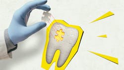
Red-complex bacteria
There are three red-complex bacteria in the mouth—Porphyromonas gingivalis, Treponema denticola, and Tannerella forsythia1—which are considered a root cause of periodontal disease. The level of destruction depends on the host response to the bacteria, the patient’s diet, and home care. The systemic burden determines how quickly these bacteria can replicate and cause destruction. A. actinomycetemcomitans and P. gingivalis can transfer their high virulence factors to other bacteria in the mouth. P. gingivalis can induce dysbiotic gut microbiota to reproduce gut pathology. Scaling and root planing alone will not eliminate the entire load of red-complex bacteria.2
You may also be interested in ... Dental abscesses and their systemic effects
Once we identify the type of red-complex bacteria, we can recommend adjunctive therapies to eliminate or reduce the pathogenic loads. For example, xylitol can be used to kill P. gingivalis.3 Multiple tests can determine the red-complex bacteria. I use antibiotics as a last resort, so it’s advantageous to select a test that provides sensitive measurements. Tests range from measuring red complex at the thousand counts to the 10 counts of bacteria.
pH testing
The critical pH of the mouth is 5.5 on enamel and 4.5 on cementum. Many sugar-free beverages are below the critical pH, including bottled water. In a healthy mouth, the saliva can buffer that pH back up to a more alkaline level; however, when the patient is experiencing even mild levels of xerostomia, this ability is compromised.
Over the last decade, we’ve educated our patients to avoid sugary beverages. Despite being sugar free, many beverages (such as seltzer water) have a pH as acidic as orange juice. Limiting the frequency, duration, and consumption of acidic beverages will reduce the incidence of dysbiosis. Furthermore, patients with gram-negative bacteria tend to have a more acidic baseline pH.4
You may also be interested in ... Using evidence-based science to drive periodontal treatment
Implementing a pH test chairside can be as simple as using an oral pH strip before preventive hygiene therapy. Instruct the patient not to eat or drink one hour before the appointment to establish their oral pH. A salivary buffer kit can be used to test the pH, resting salivary flow, and stimulated salivary flow, which is a more comprehensive approach to testing the buffering capacity of the patient’s saliva. With inadequate buffering capacity, the patient is at higher risk for hard and soft tissue diseases, including white spot lesions.5
Sleep apnea
Sleep is designed to help detoxify the body. When the patient has sleep apnea, they struggle to have adequate oxygen, which can be life-threatening. Nearly 25% of patients aged 30-70 have some form of sleep apnea.6 A simple way to screen for sleep apnea is in our EIO assessment, using the Mallampati score:
- Class 1: Soft palate, uvula, faucial pillars fully visible
- Class 2: Soft palate, uvula, fauces visible
- Class 3: Soft palate, base of uvula visible
- Class 4: Only the hard palate is visible
A higher Mallampati score is a predictor of the risk for obstructive sleep apnea.
The complete integration of a root-cause hygiene model is more complex than a three-part system. Beginning to implement these three screenings can offer eye-opening realities of the underlying causes of dental dysbiosis. Once we identify the root cause of oral disease, we can effectively partner with and empower patients to implement technologies to support symbiosis.
Editor's note: This article appeared in the August 2023 print edition of RDH magazine. Dental hygienists in North America are eligible for a complimentary print subscription. Sign up here.
References
- Suzuki N, Yoneda M, Hirofuji T. Mixed red-complex bacterial infection in periodontitis. Int J Dent. 2013;2013:587279. doi:10.1155/2013/587279
- Liu G, Luan Q, Chen F, Chen Z, Zhang Q, Yu X. Shift in the subgingival microbiome following scaling and root planing in generalized aggressive periodontitis. J Clin Periodontol. 2018;45(4):440-452. doi:10.1111/jcpe.12862
- Han SJ, Jeong SY, Nam YJ, Yang KH, Lim HS, Chung J. Xylitol inhibits inflammatory cytokine expression induced by lipopolysaccharide from Porphyromonas gingivalis. Clin Diagn Lab Immunol. 2005;12(11):1285-1291. doi:10.1128/CDLI.12.11.1285-1291.2005
- Kilian M, Chapple ILC, Hannig M, et al. The oral microbiome – an update for oral healthcare professionals. Br Dent J. 2016;221(10):657-666. doi:10.1038/sj.bdj.2016.865
- Bechir F, Pacurar M, Tohati A, Bataga SM. Comparative study of salivary pH, buffer capacity, and flow in patients with and without gastroesophageal reflux disease. Int J Environ Res Public Health. 2021;19(1):201. doi:10.3390/ijerph19010201
- Finkel KJ, Searleman AC, Tymkew H, et al. Prevalence of undiagnosed obstructive sleep apnea among adult surgical patients in an academic medical center. Sleep Med. 2009;10(7):753-758. doi:10.1016/j.sleep.2008.08.007
About the Author

Amber Auger, MPH, RDH
Amber is a dental hygiene clinician, international speaker, and hygiene director who specializes in nonsurgical periodontal therapy, heart-centered education, and efficiency. Through her signature program, Thrive in the OP and Thrive Chairside Summit, she equips hygienists with evidence-based systems to elevate patient outcomes, confidence, and production. Amber blends clinical expertise with practical strategies to help dental teams implement sustainable, science-driven protocols. She can be reached at amberauger.com.