Implication of brittle bone diseases
Osteogenesis has been found in an Egyptian infant mummy from about 1000 BC. Although it is not a new disease, it is a rare disease. Since it has some dental commonalities, dental professionals should be able to recognize the disorder and discuss dental options intelligently with the patient, family, and other health-care providers.
Osteopenia and bone fragility are characteristics of the bone disease called osteogenesis imperfecta (OI). It is the most commonly inherited bone disease and is classified as a metabolic connective tissue synthesis disorder. The Type 1 collagen that is needed for connective tissue is reduced and of poor quality throughout the body.
According to the Osteogenesis Imperfecta Foundation, OI is lifelong and rare, affecting approximately 25,000 to 50,000 people in the United States. OI manifest in varying types ranging from mild to severe classifications—Type l through Type lV with subclassifications of over 15 types with variations as well.
Also by Nancy Burkhart
Macroglossia and the disease implications
Is your patient juuling?
Type 1 is milder with minimal bone deformity. Broken bones from very little trauma are usually the first indication of OI. The individual reaches normal stature, but it does affect the bones causing some breakage/fractures, hearing loss, blue sclera (whites of the eyes have blue, grey or purple tint), and the teeth may or may not be affected.
Type ll is seen in utero, very lethal, and the infant usually suffers respiratory distress early. Infants with osteogenesis imperfecta may present with shortened legs and arms and simply turning the baby over may break a bone. There may be bowing of the legs and curvature of the spine, and frontal bossing in some cases with OI.
When the teeth present with enamel loss, cracking and discoloration the term dentinogenesis imperfecta is usually diagnosed. The defective dentin does not support the enamel long-term.
Type lll is severe with all characteristics noted but in a very deforming manner. Type lV has similar characteristics but does involve the sclera and the teeth. The patient presented in this case study had her first broken bone at 3 months old and is now an adult. She has suffered over 24 broken bones over her lifetime, but leads a normal life.
Since there is a hereditary component involved in osteogenesis imperfecta, patients may already have a known relative that may be documented as having the disorder. Depending upon the type of OI, the gene is passed by parents, or there may be a distant relative who also carried the gene. OI is an autosomal dominant, autosomal recessive disease or may be noted as a sporadic mutation. In most cases, 90% or more, OI is caused by a faulty gene and affects either the amount, quality and/or type of collagen that is produced throughout the body. The remaining 10% are caused by inherited genes that are mutated in a recessive or dominant pattern.
The OI Foundation recommends that siblings be tested since they have a 67% of being a carrier of the recessive gene.
The bones are affected with synthesis defects and all skeletal aspects, causing bowing of the legs, curvature of the spine, cranial abnormality, tendons & ligaments are affected, as well as the skin. Many oral manifestations are evident as well. Ocular and dental characteristics of this disorder are discussed below. Osteogenesis Imperfecta should not be confused with dentinogenesis imperfecta since they result from different mutations and vary in characteristics for each. Many patients with dentinogenesis imperfecta will also prove to have osteogenesis imperfecta when tested (Kessler, 2013) (Teixeira,2008).
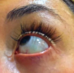
Ocular involvement—Ocular involvement (see Figure 1) is usually most evident extraorally. Even before the dental professional begins an oral exam, the eyes may be noticed in an extraoral exam as having a bluish, grey, or purple tint. The effects on the eyes may include blue sclera, glaucoma, myopia and keratoconus (progressive thinning of the cornea).
The blue sclera is sometimes very subtle and may be more noticeable if a white sheet of paper is held near the eye for color comparison. The sclera is composed of Type 1 collagen and develops in OI much thinner than in normal ocular tissue.
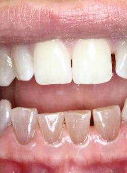
Clinical features—Discoloration (see Figure 2) is noticed more in the primary teeth than in the permanent teeth of those with osteogenesis imperfecta. The teeth have a brown, yellow to gray color. The teeth presented in this image are all natural teeth with the maxillary teeth appearing clinically normal and bright white. This is typical in OI; some teeth may appear normal in color.
There are significant oral manifestations that will vary in the effects on the teeth, the pulp ... The teeth chip and break easily and are often misaligned. ... there is a decrease in vertical dimension and the possibility of tooth loss.
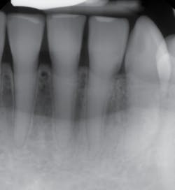
Oral ramifications affect the dentin with color variations, and this is evident in the mandibular teeth (see Figure 3). There are significant oral manifestations that will vary in the effects on the teeth, the pulp, and the overall oral clinical presentation of the patient.
The teeth chip and break easily and are often misaligned. Because of tooth structure loss/attrition, there is a decrease in vertical dimension and the possibility of tooth loss. Often referred to as “dentinogenesis imperfecta” when seen clinically, the two are separate entities even though the effects on the teeth are visually identical. When this clinical appearance occurs with the teeth, it is usually termed dentinogenesis imperfecta, and the patient may or may not have osteogenesis imperfecta, but many patients do and this may be subsequently diagnosed with testing.
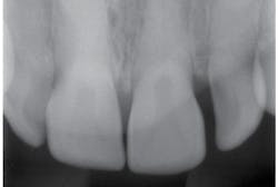
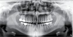
Radiographs—The teeth exhibit pulpal-root canal obliteration (see Figures 4, 5). The roots of the teeth are narrow and appear to be funnel-shaped. The crowns are large and there is constriction at the cervical neck of the tooth.
Treatment—Reconstruction of the teeth is often needed for patients who have extensive destruction. The images presented in this article show that clinically the maxillary teeth are esthetically pleasing and even bright white. The mandibular teeth do exhibit the typical brown opalescent teeth that are seen with this type of OI.
Bisphosphonate treatment is a commonly used therapy in certain cases of OI to stabilize bone and prevent fractures. The side-effect could be medication-related osteonecrosis of the jaw (MRONJ). Implants may be utilized for tooth replacement and the success is varied. Treatment is palliative but not curative; and the goal is the achievement of life-long health.
Dental professionals play a large role in monitoring and initially assessing the medical/dental status of the patient. These patients need a wide array of health-care providers to proceed with full treatment and long-term health. Dental professionals should understand disease processes and be able to meet the needs of medically compromised patients.
As always, listen to your patients and continue to ask good questions.
Originally published in 2018 and updated regularly.
References
1. Teixeira CS, Santos Felippe MC; Felippe WT, Silva-Sousa YT; Sousa-Neto MD. The role of dentists in diagnosing osteogenesis imperfecta in patient with dentinogenesis imperfecta. JADA, July, (7)2008 139: 906-914.
2. Kessler H. De Long L, Burkhart NW. General & Oral Pathology for The Dental Hygienists. 2nd edition, 2013. Lippincott, Williams & Wilkins & Page 575.
3. Milano M, Wright T, Loechner K. Dental implications of osteogenesis imperfecta: Treatment with lV Bisphosphnate: Pediatric 2011; Dent 33:4: 349-352.
4. Osteogenesis Imperfecta Foundation. oif.org
About the Author
Nancy W. Burkhart, EdD, MEd, BSDH, AAFAAOM
NANCY W. BURKHART, EdD, MEd, BSDH, AAFAAOM, is an adjunct professor in the Department of Periodontics-Stomatology, College of Dentistry, Texas A&M University, Dallas, Texas. She is founder and cohost of the International Oral Lichen Planus Support Group (dentistry.tamhsc.edu/olp/) and coauthor of General and Oral Pathology for the Dental Hygienist, now in its third edition. Dr. Burkhart is an academic affiliate fellow with the American Academy of Oral Medicine, where she also serves as chair of the Affiliate Fellowship Program Committee. She was awarded the Dental Professional of the Year in 2017 through the International Pemphigus and Pemphigoid Foundation, and she is a 2017 Sunstar/RDH Award of Distinction recipient. Her professional interests are in the areas of oral medicine and the relationship between oral and whole-body health, with a focus on mucosal disease and early oral cancer detection. Contact her at [email protected].
