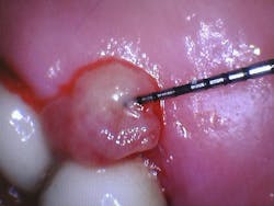
Case study: Pyogenic granuloma localized on lingual palatal mucosa of nos. 14-15 in a 72-year-old male
This case report describes an unusual presentation of a pyogenic granuloma in a 72-year-old male. Pyogenic granulomas, while common benign vascular lesions, are typically found in younger individuals and pregnant women. Their occurrence in older males underscores the importance of considering a broad differential diagnosis when encountering oral lesions.
Pyogenic granuloma: An overview
Pyogenic granuloma, though the name suggests an infectious process, is a benign vascular tumor characterized by excessive growth of capillaries and fibroblasts. While its exact etiology remains elusive, the pathogenesis is often linked to a combination of factors, including local irritation, trauma, hormonal fluctuations, and certain medications. The interaction of these factors triggers a cascade of events that leads to the characteristic clinical presentation of this lesion.1
Local irritation or trauma, such as chronic inflammation from poor oral hygiene or accidental injury, can initiate an inflammatory response that stimulates the release of growth factors and cytokines, promoting angiogenesis (new blood vessel formation) and fibroblast proliferation.2
Hormonal changes, particularly during pregnancy or puberty, can also influence the development of pyogenic granulomas due to their effects on vascular reactivity and tissue growth. Certain medications, such as oral contraceptives and retinoids, have been implicated in some cases, though the exact mechanism is unclear.
The rapid proliferation of capillaries and fibroblasts results in a highly vascularized, friable lesion that bleeds easily. This explains its clinical presentation of a solitary, red-to-purple nodule, often pedunculated or sessile, and prone to bleeding upon even slight manipulation.
These lesions can vary significantly in size, ranging from a few millimeters to several centimeters, and they typically occur on the gingiva, lips, tongue, and buccal mucosa.3 However, they can also manifest in extraoral locations, such as the skin and nasal septum.
Understanding the pathogenesis of pyogenic granuloma is crucial for accurate diagnosis and appropriate management.4 Recognizing the potential triggers and the resulting cellular changes allows clinicians to differentiate this lesion from other conditions with similar clinical presentations, such as peripheral giant cell granuloma, peripheral ossifying fibroma, hemangioma, and oral squamous cell carcinoma.4
Case presentation
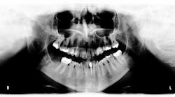
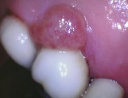
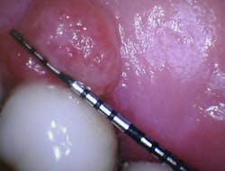
During a routine hygiene appointment, a 72-year-old man reported a "growth" on the roof of his mouth behind his upper left molar. He noted its presence for about two weeks with occasional bleeding. An oral examination revealed a solitary, reddish nodule measuring 7 mm in diameter in the gingival mucosa between teeth nos. 14 and 15.
The lesion was soft, tender to the touch, asymptomatic, and bled slightly when examined. Additionally, a defective crown on tooth no. 14 was observed. The patient indicated that the only recent change in his oral hygiene routine was using a different size of interdental brush in that area.
Diagnosis and management
An excisional biopsy was performed, and a histopathological examination confirmed the diagnosis of pyogenic granuloma. The patient was given oral hygiene instructions that focused on gentle brushing and flossing, and advised to use a smaller interdental brush in the affected area.
Discussion and conclusion
This case underscores a crucial point in clinical practice: even seemingly straightforward oral lesions can present in atypical ways. Although uncommon, the occurrence of pyogenic granuloma in an older male patient highlights the necessity of maintaining a broad differential diagnosis. While the clinical presentation was suggestive, the patient's age and the lesion's location prompted a biopsy to exclude other possibilities, such as oral squamous cell carcinoma.
This case highlights how important it is for dental professionals to take a comprehensive approach when evaluating oral lesions. Always remember to gather a detailed patient history and conduct a thorough clinical examination, even when a lesion seems familiar and inoffensive. This will ensure we can accurately identify any potential issues and intervene promptly. Early detection and proper management can truly make a difference in preventing complications and improving our patient’s overall health. Dental hygienists play a vital role in this process!
By carefully observing and raising concerns about any unusual oral findings, we actively contribute to early diagnosis and improved patient care. We have the power to make a real impact!
References
1.Lomeli Martinez SM, Carrillo Contreras NG, Gómez Sandoval JR, et al. Oral pyogenic granuloma: a narrative review. Int J Molecular Sci. 2023;4(23):16885. doi:10.3390/ijms242316885
2. Ribeiro JL, Moraes RM, Carvalho BFC, Nascimento AO, Milhan NVM, Anbinder AL. Oral pyogenic granuloma: an 18-year retrospective clinicopathological and immunohistochemical study. J Cutaneous Path. 2021;48(7):863-869. doi:10.1111/cup.13970
3. Sonar PR, Panchbhai AS. Pyogenic granuloma in the mandibular anterior gingiva: a case study. Cureus. 2024;16(1):e52273. doi:10.7759/cureus.52273
4. Ramón Ramirez J, Seoane J, Montero J, Esparza Gómez GC, Cerero R. Isolated gingival metastasis from hepatocellular carcinoma mimicking a pyogenic granuloma. J Clin Perio. 2003;30(10):926-929. doi:10.1034/j.1600-051x.2003.00391.x
About the Author

Andreina Sucre, MSc, RDH
Andreina Sucre, MSc, RDH, is an international dentist, oral pathology, and oral surgery specialist practicing dental hygiene in Miami, Florida. A passionate advocate for early pathological diagnosis, she empowers colleagues through lectures focused on oral pathologies. Andreina spoke on this topic at the 2024 ADHA Annual Conference, 2023 RDH Under One Roof, and she writes about oral pathology for RDH magazine. Committed to community outreach, she educates non-native English-speaking children on oral health and actively volunteers in dental initiatives.
