Tannerella forsythia: The legend of a red complex queen, part two
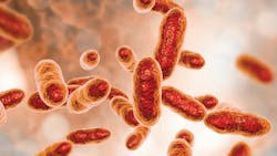
Stepping through the looking glass, we find ourselves face-to-face with the most abundant and puzzling of the red complex (RC) pathogens, Tannerella forsythia (Tf).1 Despite being linked to periodontal and endodontic infections—as well as systemic diseases such as cardiovascular disease, diabetes, and arthritis—Tf remains a mystery.2 Besides being difficult to cultivate, discoveries have exposed a period when the genomic sequence for strain FDC 92A2 was accidentally mismatched and applied to the conventional strain ATCC 43037, muddying years of research.2,3
Part one of this series reviewed how Tf leans into the Black Queen hypothesis by exchanging genes likely for energy-preserving advantages. Part two reveals how Tf thrives using the art of illusion and evasion. Known as the Red Queenhypothesis, outsmarting your opponent requires studying their patterns and adapting to changes while they do the same. Regarding Tf, an expert in manipulation, we’re up against a grandmaster. It’s Alice vs. the Red Queen in Wonderland, but in the operatory it’s clinician vs. pathogen—and every move changes the game entirely.
Life on the outer rim
Biofilms offer refuge, and not everyone’s invited. In the relatively protected region of the gingival crevice, pathogens form well-orchestrated polymicrobial biofilms based on synergy.1,3 There is intent behind the timing and order of the arrangement. Complex parameters related to nutrients, oxygen levels, resident species, and host defenses must be considered before a bacterium colonizes. For example, Fusobacterium nucleatum (Fn), a middle-colonizer, and Tf form a tight interspecies partnership through shared sialic acid sugars.
At the biofilm’s exterior, the two remaining RC members, Porphyromonas gingivalis (Pg) and Treponema denticola (Td), are rarely found without Tf.1 In fact, in 2010, Zijnge et al. pointed to Tf’s location in the “intermediate layer of subgingival plaque, whereas P. gingivalis tended to be found in micro-colonies in the top layer, and Treponemes were found outside the top layer.”1 This suggests that T. forsythia is an “early” late-colonizer and may be a “precursor species for colonization” by Pg and Td.1 Furthermore, growth and pathogenicity of T. forsythia are enhanced in the presence of other bacteria, making it an appealing ally.2 Together at the war table, the RC devise their rise to power, emerging as a collective and formidable pseudopathogen.
The S-layer
Unfortunately, arriving last to the biofilm means being the first to encounter host offenses. Readying troops for combat, T. forsythia has concocted plans to invade epithelial cells, impair polymorphonuclear leukocyte functions, and promote dysregulated inflammation.4 Again, the cell wall, and specifically the outer membrane, becomes the focus. Both Pg and Tf, members of the phylum Bacterioidetes, rely on a type 9 protein secretion system (T9SS) to export an arsenal of virulence factors to the outer membrane.4 For virtually all archaea and several bacteria, one of these measures is a reinforced surface layer, or S-layer.5
Gram-negative bacteria anchor their S-layers to the lipopolysaccharide (LPS)), using it as another protective shield to fortify against antimicrobial agents and host degradation.2,5 Generally, S-layers are crystallized monolayers composed of single glycoproteins (sugars bonded to proteins).5 “Contradicting the so far valid building plan” of S-layers, Tf covers its exterior with a uniquely zippered assemblage of two glycoproteins: TfsA and TfsB.3,5 Even rarer, the glycan (sugar) portions of the glycoprotein are O-linked (attached to the oxygen atom of serine or threonine residues on proteins) in a process called glycosylation.3 Once thought to be the only gram-negative with a glycosylated S-layer, Tf now shares this category with others such as the pro-inflammatory and middle-colonizing Campylobacter rectus (Cr).5
Illusion and evasion
Concealing the fortress are elaborate gardens and hedgerows. The goal of a periodontal pathogen is not to cause periodontal disease. Rather, a pathogen needs to create just enough inflammation to obtain sanctuary and nutrients while going unnoticed. Tf alters the S-layer glycans, adjusting their functions and environmental interactions. For camouflage, the pathogen mimics the sialic acid-covered host cells by decorating the terminal ends of the glycans with sialic acidlike residues.5 The glycan modifications provide enough disguise to regulate dendritic cell recognition, suppress Th17-mediated neutrophil infiltration, and delay cytokine responses.5
RC pathogens employ sialidases (enzymes) to harvest sialic acids as nutrients. Once cleaved, the previously hidden binding-epitopes are exposed, becoming opportunities for bacterial adhesion and invasion.4 Another shared RC virulence factor is a family of hooklike leucine-rich repeat proteins (LRRs) known for their robust attachment.1 For example, Td uses its LRR (LrrA) to bind to Tf.1 Tf’s LRR is the bacterial surface protein antigen, BspA, and doubles as a surface-secreted glycoprotein acting like a skeleton key providing adhesion and entry into cells.2 BspA triggers “the production of IL-8,” a chemokine produced by macrophages, and in this example, epithelial cells, “through a TLR2 dependent mechanism,” releasing additional nutritional sources.4
Proteases
To a hygienist, disease is a battlefield full of bleeding, bone loss, and recession. To an RC pathogen, it’s the day-after victory celebrations from the feast in the great hall. The skilled swordsmen for pathogens are proteases, enzymes specializing in destruction. Pg uses gingipains, Td uses dentipains, and Prevotella intermedia uses interpains. Tf’s proteases, however, are still being investigated. For now, “various proteases have been implicated in T. forsythia’s pathogenicity, including, e.g., PrtH proteases which are associated with attachment loss, a trypsin-like cysteine protease … and secretory KLIKK proteases capable of targeting … collagen, gelatine, elastin and casein.”4
Outer membrane vesicles
Archers can guarantee success in battle; targeting the enemy from great distances limits physical interactions. Outer membrane vesicles (OMVs) are “emerging as ‘bacterial warfare’ agents in the pathogenesis of periodontitis.”3 Increasing numbers of gram-negative bacteria are found to secrete spherical particles budding off from the OM, including all three of the RC.3 Miniaturized, OMVs travel farther into the environment and deeper into cells or tissues, delivering their toxic cargo.3
Remarkably, OMVs are packaged with virulence factors similar to the main pathogen. For Tf, this includes: genes for a T9SS, an intact S-layer enriched with TfsA and TfsB glycoproteins, the surface antigen BspA, and several of the known Tf-specific proteases. “The response of host cells to T. forsythia OMVs is comparable to or even stronger than that elicited by the whole bacterium.”4 Tf’s OMV virulence induces “the production of TNF-α and IL-8 in macrophages, and IL-6, IL-8, and MCP-1 in human periodontal ligament mesenchymal stromal cells.”4
Reflections
We are far from fully understanding the complexities of this ancient adversary. Still, the mirror reflects the true journey: hygienists working together to solve the riddles of oral pathogens.
Editor's note: This article appeared in the March 2026 print edition of RDH magazine. Dental hygienists in North America are eligible for a complimentary print subscription. Sign up here.
References:
-
Dashper SG, Seers CA, Tan KH, Reynolds EC. Virulence factors of the oral spirochete Treponema denticola. J Dent Res. 2011;90(6):691-703. doi:10.1177/0022034510385242
-
Amano A, Chen C, Honma K, et al. Genetic characteristics and pathogenic mechanisms of periodontal pathogens. Adv Dent Res. 2014;26(1):15-22. doi:10.1177/0022034514526237
-
Friedrich V, Gruber C, Nimeth I, et al. Outer membrane vesicles of Tannerella forsythia: biogenesis, composition, and virulence. Mol Oral Microbiol. 2015;30(6):451-473. doi:10.1111/omi.12104
-
Schäffer C, Andrukhov O. The intriguing strategies of Tannerella forsythia’s host interaction. Front Oral Health. 2024;5:1434217. doi:10.3389/froh.2024.1434217
-
Posch G, Sekot G, Friedrich V, et al. Glycobiology aspects of the periodontal pathogen Tannerella forsythia. Biomolecules. 2012;2(4):467-482. doi:10.3390/biom2040467
About the Author

Holly Moons, CRDH
Holly has devoted 25 years to periodontal dental hygiene. She has served in multiple roles on local and state boards, belongs to several study clubs, and continues to publish and speak nationally. Holly advocates for a collaborative dental-medical model focused on reducing chronic diseases. Her passion for microbiology drives her research on polymicrobial synergy, pathogenic virulence factors, and emerging concepts in biofilm expression. Connect with her at [email protected].